
Lahalak Medical PlatformDifferentiate Care
Learn when a mole or skin lesion requires a biopsy. Understand the ABCDE criteria, biopsy types, procedure steps, and when to see a dermatologist.

Advertisement
The skin is our body's primary shield against external elements, and moles (nevi) are common skin growths that most people develop over time. In the vast majority of cases, these moles are entirely benign and pose no health risks. However, abnormal changes can sometimes occur in existing moles, or new, suspicious skin lesions may appear. In such cases, a skin biopsy serves as a critical diagnostic tool to evaluate skin cells and rule out skin cancers, particularly melanoma. This comprehensive guide details when a mole or skin lesion requires a biopsy, what the procedure entails, and the key warning signs to watch for.
A skin biopsy is a straightforward, safe medical procedure typically performed in a dermatologist's clinic under local anesthesia. During the procedure, the doctor removes a small sample of skin cells or the entire suspicious lesion. This sample is then sent to a laboratory for histopathological analysis under a microscope by a dermatopathologist. This precise examination determines whether the cells are normal, benign, atypical, or cancerous, allowing for an accurate and early treatment plan.
To help individuals identify moles that may require medical evaluation, dermatologists developed the widely recognized ABCDE criteria. This framework is highly effective for performing a regular self-examination of moles to detect early changes:
In addition to the ABCDE rule, there are other clinical signs that should be taken seriously, including:
Biopsies are not limited to moles; they are performed on various skin lesions that exhibit suspicious clinical features. These include:
These are benign moles that look unusual under a microscope and can clinically mimic melanoma. Individuals with numerous atypical nevi have a higher risk of developing skin cancer, making regular monitoring and selective biopsies essential.
The most common type of skin cancer, BCC typically develops on sun-exposed areas like the face and neck. It often appears as a pearly or pink bump, a flat scar-like lesion, or a sore that bleeds easily and fails to heal completely.
Arising in the squamous cells of the outer skin layers, SCC often presents as a firm red nodule, a rough scaly patch, or an open sore. A biopsy is crucial to diagnose SCC early and prevent it from spreading to deeper tissues.
The most serious form of skin cancer, melanoma originates in the pigment-producing melanocytes. It has a high potential to spread rapidly to other organs if not diagnosed and treated early. An excisional biopsy is the gold standard for diagnosing melanoma and determining its depth of invasion.
The choice of biopsy technique depends on the size, location, and depth of the suspected lesion. The following table compares the primary types of skin biopsies:
| Biopsy Type | Procedure Method | Common Indications | Approximate Healing Time |
|---|---|---|---|
| Shave Biopsy | The doctor uses a thin blade to shave off the superficial layers of the skin (epidermis and upper dermis). | Superficial skin lesions, raised moles, and actinic keratoses. | 7 to 10 days (usually requires no stitches). |
| Punch Biopsy | A circular, pen-like tool is used to remove a deeper core of skin, including the deeper dermis and subcutaneous fat. | Deep skin lesions, unexplained rashes, and small moles. | 10 to 14 days (may require one or two stitches). |
| Excisional Biopsy | The entire skin lesion is removed along with a small margin of surrounding healthy skin using a scalpel. | Moles suspected of being melanoma and larger skin tumors. | 2 to 3 weeks (requires surgical sutures). |
| Incisional Biopsy | Only a portion or wedge of a large skin lesion is removed for diagnosis before planning definitive removal. | Very large tumors or lesions in cosmetically sensitive areas like the face. | 10 to 14 days (requires surgical sutures). |
Understanding the steps of a skin biopsy can significantly reduce patient anxiety. Here is what to expect before, during, and after the procedure:
A skin biopsy requires minimal preparation. You must inform your doctor if you have any allergies (especially to local anesthetics like lidocaine), if you take blood-thinning medications (such as aspirin or warfarin), or if you have a history of bleeding disorders or poor wound healing.
Proper home care is vital for rapid healing and minimizing scar formation. Key instructions include:
Biopsy results are typically ready within several days to two weeks. When reviewing the pathology report with your doctor, the findings will generally fall into one of these categories:
Given the hot, sunny climate in Saudi Arabia, prolonged exposure to harmful ultraviolet (UV) radiation is a major risk factor for skin changes. Therefore, it is highly recommended to schedule an annual dermatologist consultation for a comprehensive full-body skin check, especially if you have a family history of skin cancer, fair skin, or numerous moles. Remember, early detection of skin changes increases cure rates to nearly 100% for many lesions, preventing complex health complications.
Clear, direct answers
Advertisement
Continue exploring
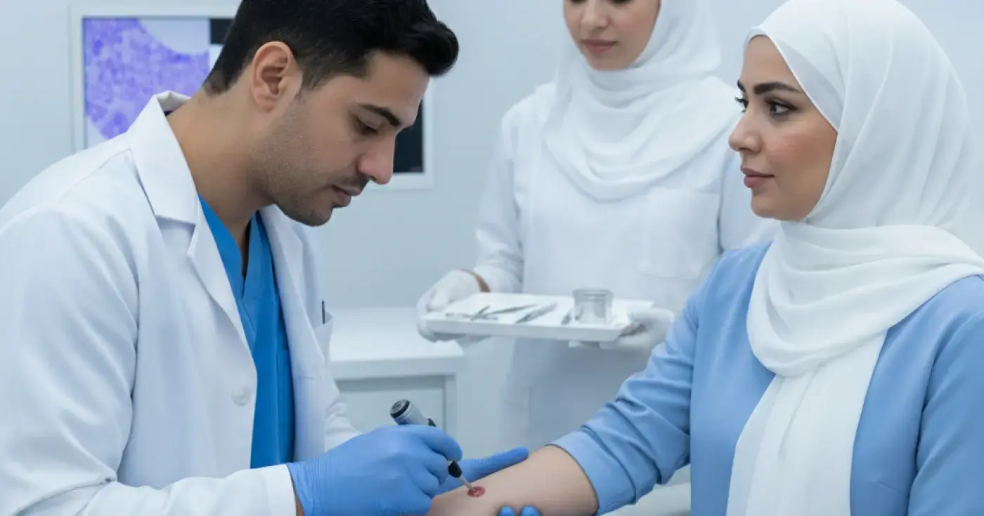
Learn about the different types of skin biopsies (shave, punch, excisional), their uses, preparation, recovery, and how to care for your wound.
Read article
A comprehensive medical guide on how a skin biopsy is performed, its types, preparation steps, wound care, and understanding your results.
Read article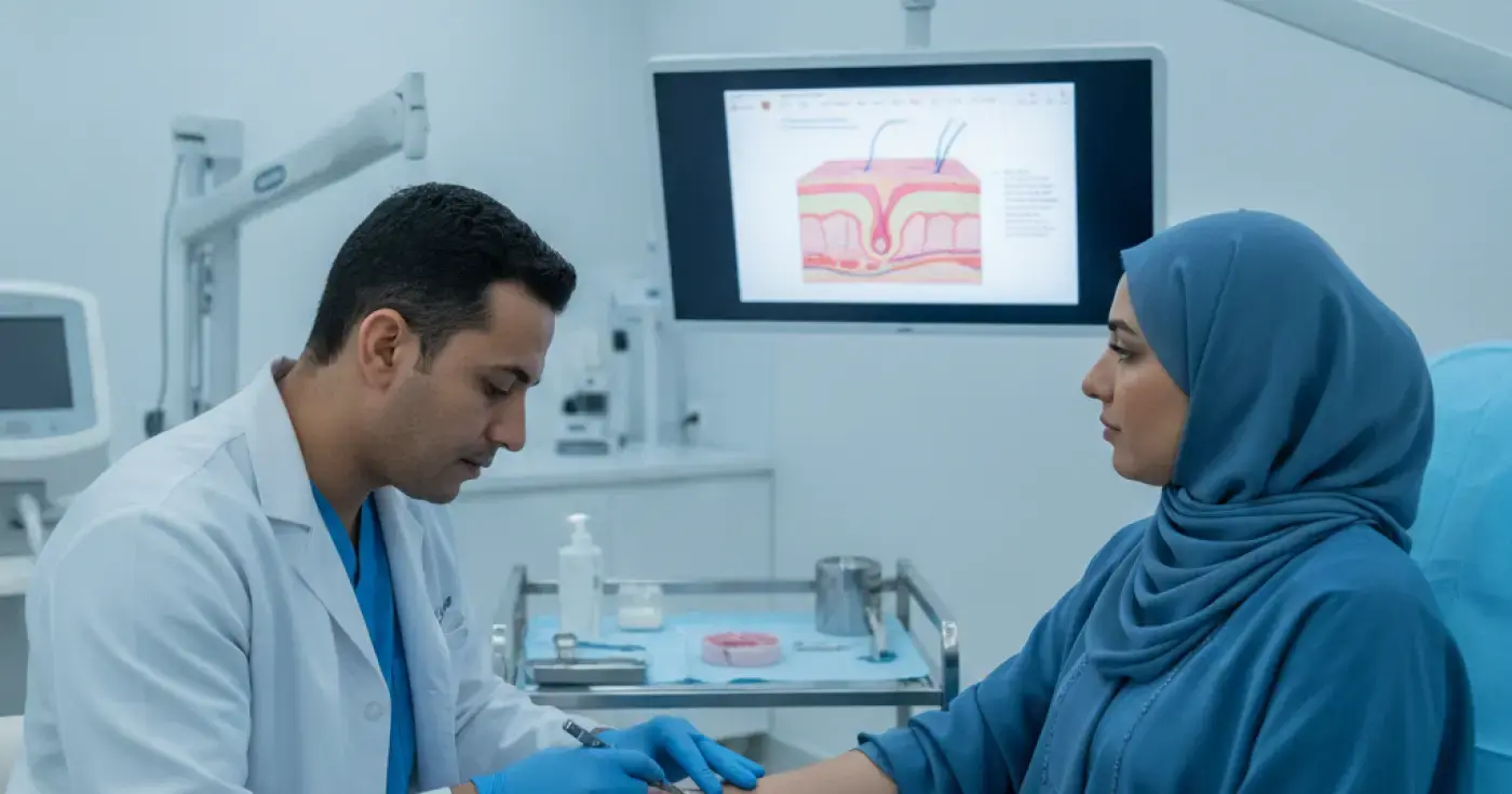
Learn what a skin biopsy is, why doctors recommend it, the different types (shave, punch, excision), and how to care for your wound post-procedure.
Read articleExplore the topic