
Lahalak Medical PlatformDifferentiate Care
Learn about the different types of skin biopsies (shave, punch, excisional), their uses, preparation, recovery, and how to care for your wound.
Advertisement
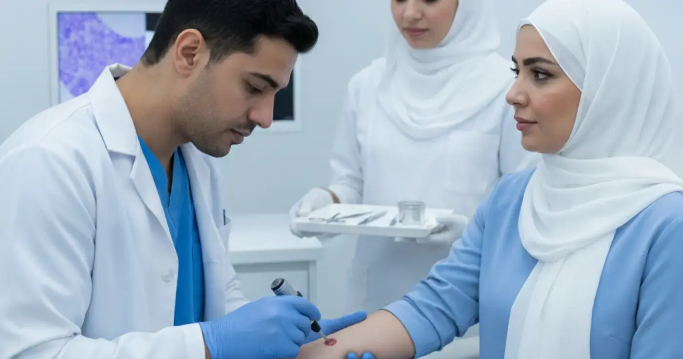
The skin is the body's first line of defense and is prone to various changes and lesions over time. Often, a visual examination with the naked eye or even with a dermoscope is not enough to reach a definitive diagnosis. This is where a skin biopsy comes in as an essential, safe medical procedure in which a dermatologist removes a small sample of skin cells or tissue for histopathological analysis under a microscope.
The clinical importance of a skin biopsy lies in its ability to differentiate between benign, precancerous, and malignant skin lesions, as well as to diagnose chronic inflammatory skin conditions such as psoriasis, eczema, cutaneous lupus erythematosus, and deep fungal or bacterial infections. This highly accurate procedure helps physicians design a personalized and effective treatment plan based on confirmed laboratory results.
A dermatologist decides to perform a biopsy when there are doubts about the nature of a specific skin lesion, or when a skin condition does not respond to conventional treatments. Key medical reasons for performing a biopsy include:
Skin biopsy types vary based on the size, location, and depth of the skin lesion required to reach an accurate diagnosis. The physician selects the most appropriate technique for each individual case. Here are the details of the four main types:
A shave biopsy is one of the most common and least invasive types. During this procedure, the doctor uses a small, sharp, flexible blade or a scalpel to shave off the superficial layers of the skin (the epidermis and the upper part of the dermis). This is typically performed for elevated skin lesions that project above the skin surface, such as skin tags, seborrheic keratoses, and certain benign moles. A major advantage of this procedure is that it does not require sutures (stitches), heals quickly, and leaves a minimal scar.
A punch biopsy is used to obtain a deeper sample that includes all three layers of the skin: the epidermis, dermis, and subcutaneous (fatty) tissue. The physician uses a sharp, circular tool resembling a tiny cookie cutter, usually ranging from 2 to 6 millimeters in diameter. The tool is gently rotated to cut a cylinder of skin. This type is ideal for diagnosing inflammatory skin rashes, autoimmune diseases, and lesions extending deep into the skin. Depending on the size of the punch, the wound may require one or two sutures to facilitate rapid healing and minimize scarring.
In an excisional biopsy, the doctor removes the entire skin lesion along with a small margin of surrounding healthy skin to ensure complete clearance. A traditional surgical scalpel is used to make an elliptical cut down to the deep fatty tissue. This procedure is primarily used when malignant tumors like melanoma are suspected, or when removing a subcutaneous lump such as a sebaceous cyst or lipoma. This procedure always requires closing the wound with cosmetic sutures.
An incisional biopsy is similar to an excisional biopsy, but the key difference is that only a portion (a sample) of a very large or critically located skin mass is removed. This is performed to confirm the diagnosis first before planning a major surgical procedure for full removal. This procedure also requires surgical sutures to close the wound.
The following table summarizes the key differences between the various types of skin biopsies for easier understanding:
| Biopsy Type | Target Depth in Skin | Tools Used | Common Indications | Need for Sutures |
|---|---|---|---|---|
| Shave Biopsy | Superficial (Epidermis & upper Dermis) | Flexible medical blade or scalpel | Skin tags, superficial moles, seborrheic keratosis | No sutures needed |
| Punch Biopsy | Deep (Epidermis, Dermis, & Fat) | Circular punch tool | Inflammatory rashes, autoimmune diseases, deep lesions | Yes (usually 1-2 stitches) |
| Excisional Biopsy | Full Depth (with safety margin) | Surgical scalpel | Suspected tumors (melanoma), sebaceous cysts | Yes (complete surgical closure) |
| Incisional Biopsy | Partial sample of a deep lesion | Surgical scalpel | Very large masses to confirm diagnosis before full excision | Yes (complete surgical closure) |
A skin biopsy is a simple, outpatient procedure performed in a dermatologist's clinic under local anesthesia, requiring minimal preparation. However, patients should follow these important guidelines to ensure safety:
A skin biopsy typically takes between 15 and 30 minutes and is performed through the following steps:
Proper wound care after a biopsy is a crucial step to ensure rapid healing and minimize the risk of infection or prominent scarring. Here are the key medical tips for daily wound care:
Although a skin biopsy is highly safe, like any minor surgical procedure, it carries some rare risks, including:
After the sample is taken, it is sent to a dermatopathology laboratory. A pathologist examines the cells under a microscope and prepares a detailed report, which usually takes from several days to two weeks. Results generally fall into:
Monitor your wound closely and contact your medical clinic immediately if you notice any of the following signs of complications:
Clear, direct answers
Advertisement
Continue exploring

A comprehensive medical guide on how a skin biopsy is performed, its types, preparation steps, wound care, and understanding your results.
Read article
Learn what a skin biopsy is, why doctors recommend it, the different types (shave, punch, excision), and how to care for your wound post-procedure.
Read article
Learn about the essential tests, biopsies, and blood work required before benign skin tumor removal to ensure safety and prevent complications.
Read articleExplore the topic