
Lahalak Medical PlatformDifferentiate Care
Discover the importance of microscopic hair and scalp analysis (trichoscopy) to diagnose hair loss causes and find the right treatment with dermatologists.
Advertisement
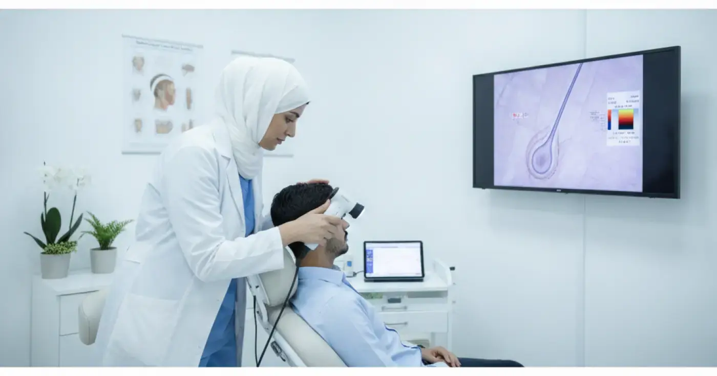
Many individuals suffer from hair loss and thinning without knowing the real cause behind it. In the past, diagnosing hair problems relied heavily on simple visual examination or manual hair pull tests. Today, medical advancements have introduced microscopic hair analysis (trichoscopy), an advanced, non-invasive imaging technique that allows dermatologists to examine the scalp and hair follicles at high magnification levels.
This precise examination helps visualize details invisible to the naked eye, such as follicular opening integrity, hair shaft thickness, scalp vascular patterns, and signs of inflammation or scaling. Through this deep insight, doctors can accurately differentiate between various types of hair loss and establish a scientifically sound treatment plan.
Identifying the underlying cause of hair problems is the first and most crucial step in the treatment journey. Random treatments or using commercial products without an accurate diagnosis can worsen the problem and waste time and money. The importance of performing a microscopic hair loss analysis lies in its ability to provide clear answers about the nature of hair loss, whether it is due to genetic factors, autoimmune disorders, localized inflammation, or nutritional deficiencies.
Additionally, microscopic analysis allows doctors to monitor the patient's response to treatment over time. By regularly comparing microscopic images taken before and after treatment, the physician can evaluate improvements in hair density and shaft thickness, adjusting the treatment plan as necessary to ensure optimal results.
Several warning signs appearing on your hair or scalp indicate the need to visit a dermatologist for a microscopic examination. Here are the most prominent signs:
Microscopic analysis is a decisive tool in diagnosing and classifying a wide range of dermatological disorders affecting the scalp and hair, including:
This condition is characterized by significant variation in hair shaft diameter (thick hairs next to extremely thin or vellus hairs) and progressive follicular miniaturization. Microscopic analysis helps detect these changes in their very early stages before thinning becomes visible to the naked eye.
An autoimmune disorder where the body's immune system attacks hair follicles. Microscopic examination reveals highly characteristic signs of alopecia areata, such as "exclamation mark hairs" and black dots within empty follicular openings.
This usually occurs after severe physical or psychological stress, childbirth, or acute illness. Microscopic analysis helps rule out other causes and confirms that a large percentage of hair has prematurely entered the resting (telogen) phase.
This is considered a medical emergency in dermatology because it leads to permanent destruction of hair follicles and their replacement with scar tissue. Microscopic analysis contributes to the early diagnosis of scarring alopecia (such as Lichen Planopilaris) to save viable follicles before permanent loss occurs.
The microscopic hair analysis procedure is simple, quick, and completely painless. It requires no anesthesia or complex preparations from the patient. Here are the typical steps performed in the clinic:
The following table illustrates the fundamental differences between modern microscopic hair analysis and traditional clinical examination:
| Comparison Feature | Traditional Clinical Examination | Microscopic Hair Analysis (Trichoscopy) |
|---|---|---|
| Tools Used | Naked eye and manual palpation | Advanced digital magnification device (Trichoscope) |
| Accuracy Level | Limited, relies only on visible surface changes | Very high, reveals microscopic changes in follicles and shafts |
| Early Diagnosis | Difficult in the early stages of the disease | Highly effective before visible thinning or bald patches appear |
| Documentation | Relies on written descriptions or standard photos | Saves high-definition magnified digital images for follow-up |
| Determining Hair Loss Type | May frequently require invasive scalp biopsies | Significantly reduces the need for painful surgical biopsies |
When a dermatologist looks through the trichoscope, they look for specific biological markers that help formulate an accurate diagnosis, including:
The diameter and consistency of the hair shaft are evaluated. Significant variation in diameter points to androgenetic alopecia. Structural abnormalities or fractures caused by excessive heat or harsh chemical treatments are also identified.
Follicular openings reflect scalp health. Yellow dots indicate sebum and keratin accumulation, while black dots represent destroyed hair shafts within the follicle (common in alopecia areata). The complete absence of follicular openings indicates scarring (cicatricial alopecia).
Examining the microvasculature helps detect active inflammation. The presence of peripilar casts or scaling may indicate inflammatory skin conditions like lichen planopilaris or psoriasis, requiring a specialized medical scalp care protocol.
Based on the microscopic findings, the doctor designs a comprehensive, personalized treatment plan. This plan may include topical treatments like minoxidil, oral medications to regulate hormones, or nutritional supplements to correct vitamin and mineral deficiencies. In advanced cases of hair loss, the doctor may guide the patient toward options like hair transplantation as a permanent solution to restore lost density.
It is essential to adhere to regular follow-up sessions (usually every 3 to 6 months) to repeat the microscopic examination and document progress, which helps adjust dosages and ensure long-term scalp health.
Given the hot and dry climate in Saudi Arabia, along with the characteristics of desalinated tap water, the scalp requires special care to prevent dryness and hair loss:
In conclusion, microscopic hair and scalp analysis represents a major leap in dermatology and aesthetics, saving time and effort in diagnosing hair disorders safely and accurately. If you suffer from persistent hair loss or notice unusual changes in your scalp, the best step is to book an appointment with a specialized dermatologist to undergo this simple test and protect your hair from future damage.
Clear, direct answers
Advertisement
Continue exploring
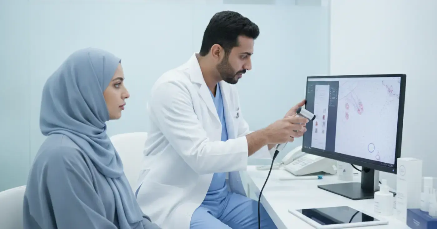
Discover how trichoscopy helps dermatologists diagnose dandruff, seborrheic dermatitis, and scalp inflammation for targeted, effective treatment.
Read article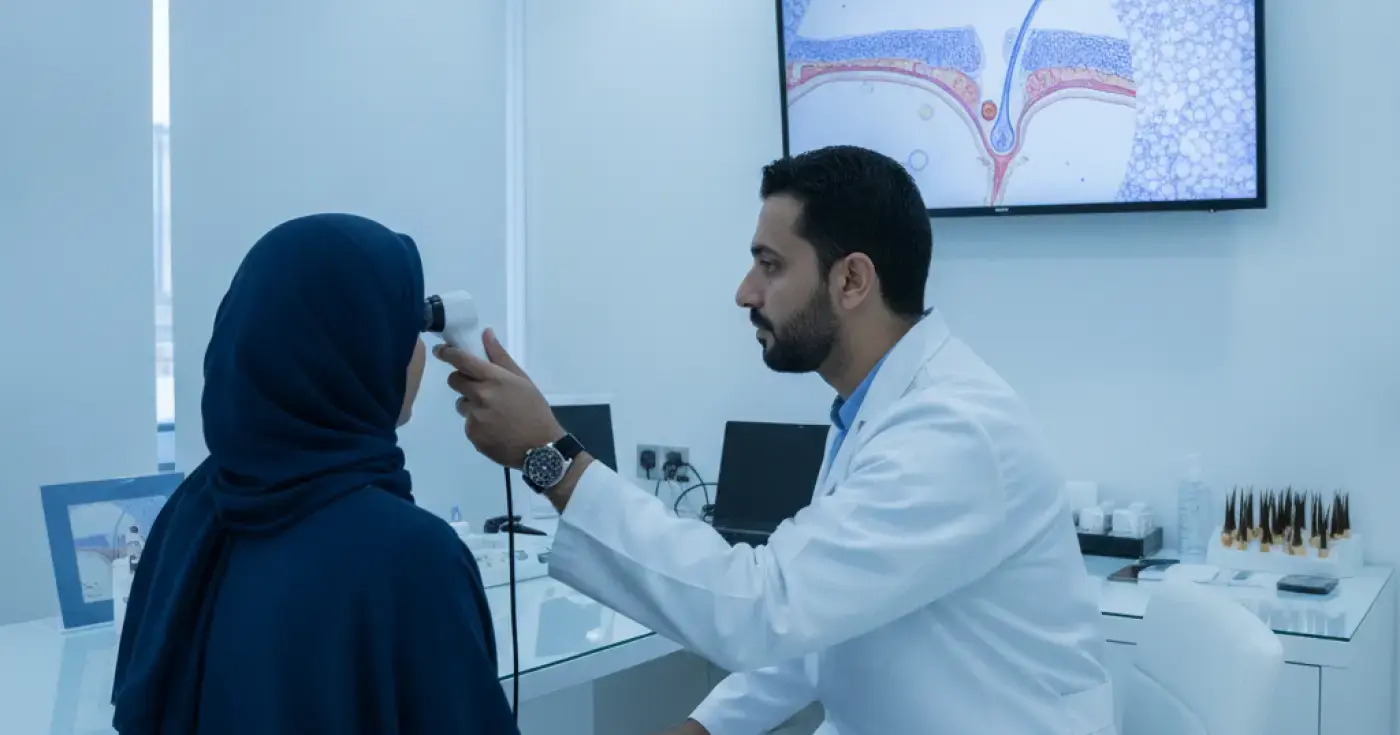
Learn how microscopic hair follicle analysis (trichoscopy) helps diagnose hair loss causes, identify scalp conditions, and guide targeted treatments.
Read article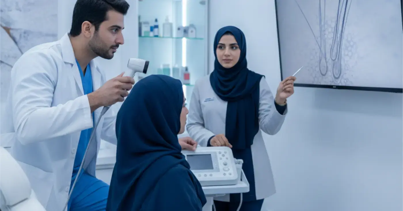
Discover how scalp trichoscopy (microscopic examination) helps dermatologists diagnose hair loss, alopecia, and scalp inflammation accurately and painlessly.
Read articleExplore the topic