
Lahalak Medical PlatformDifferentiate Care
Discover how trichoscopy helps dermatologists diagnose dandruff, seborrheic dermatitis, and scalp inflammation for targeted, effective treatment.
Advertisement
Scalp health is a fundamental pillar of healthy hair growth and overall well-being. However, a vast number of individuals suffer from chronic and bothersome scalp conditions, such as dandruff and various forms of scalp inflammation. Because these conditions often present with overlapping clinical symptoms, visual inspection with the naked eye is frequently insufficient for an accurate diagnosis. Modern dermatology addresses this challenge through advanced diagnostic tools that allow clinicians to visualize cellular and vascular details of the scalp with high precision.
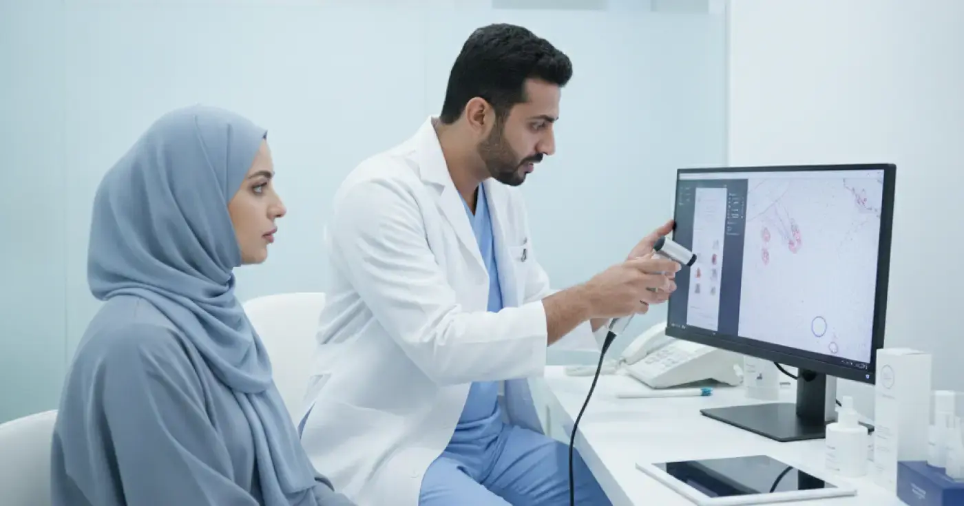
Trichoscopy is a non-invasive, state-of-the-art diagnostic technique that utilizes a handheld digital trichoscope to magnify the scalp surface and hair follicles by 10x to over 100x. This examination allows dermatologists to evaluate hair shaft structure, follicular openings, the surrounding epidermis, and microvascular patterns that are invisible to the naked eye. This quick, painless procedure is highly effective in diagnosing hair loss etiologies, scalp inflammatory disorders, and differentiating between clinically similar dermatological conditions.
A common clinical misconception is conflating simple dandruff with seborrheic dermatitis. Although both present with scaling, their underlying pathophysiology differs significantly:
During a professional microscopic hair analysis, dermatologists can pinpoint the precise factors contributing to the scalp condition, including:
1. Malassezia Yeast Proliferation: This commensal yeast plays a key role in triggering inflammation and scaling when it interacts with excess sebum. Trichoscopy reveals characteristic perifollicular scaling as an inflammatory response to yeast activity.
2. Sebum Hypersecretion: The digital trichoscope reveals oily, reflective sheets covering the scalp surface and pooling around the hair shafts, which can clog pores and foster anaerobic microbial growth.
3. Skin Barrier Dysfunction: Microscopic views can show micro-fissures and epidermal dryness, indicating a compromised skin barrier that is highly susceptible to external irritants.
Dermatologists rely on specific microscopic signs and vascular signatures to establish an accurate diagnosis, including:
A microscopic scalp examination is a straightforward, outpatient procedure consisting of the following steps:
First, the dermatologist parts the hair systematically to expose different areas of the scalp. Second, the digital trichoscope lens is placed gently against the skin, capturing high-resolution magnified images from multiple anatomical sites (such as the frontal, parietal, and occipital regions). Third, the images are analyzed in real-time on a digital monitor, comparing vascular structures, scaling patterns, and follicular health to formulate a precise diagnosis and a personalized scalp care regimen.
| Condition | Clinical Appearance (Naked Eye) | Trichoscopic Features (Magnified) | Associated Symptoms |
|---|---|---|---|
| Simple Dandruff | Small, dry, white scattered flakes | Thin, white, non-adherent scales around follicles | Mild or no itching; absence of erythema | Seborrheic Dermatitis | Thick, greasy, yellowish scales on red skin | Dotted and branching vessels; greasy perifollicular scales | Moderate to severe itching; redness; oily discharge | Scalp Psoriasis | Well-demarcated red plaques with silvery scales | Twisted, looped, ring-like vessels; dense silvery-white scales | Intense itching; extreme dryness; potential cracking/bleeding |
While many over-the-counter hair care products exist, certain clinical presentations warrant a professional dermatological evaluation and trichoscopic assessment, including:
An accurate microscopic diagnosis enables a highly targeted therapeutic protocol designed to address the root cause of the condition, which may include:
1. Topical Antifungals: Shampoos or topical solutions containing ketoconazole or ciclopirox olamine to reduce Malassezia yeast populations.
2. Topical Anti-inflammatories: For acute flares of seborrheic dermatitis or psoriasis, short-term use of topical corticosteroids may be prescribed to rapidly calm erythema and pruritus.
3. Keratolytic Agents: Formulations containing salicylic acid or urea to gently break down and exfoliate thick, adherent scales without damaging the hair follicles.
4. Sebum-Regulating Agents: Medical-grade topical treatments that help normalize sebaceous gland activity and restore a healthy scalp microbiome.
The hot and arid climate prevalent in most regions of Saudi Arabia requires specific scalp care strategies to prevent irritation and fungal overgrowth:
Clear, direct answers
Advertisement
Continue exploring
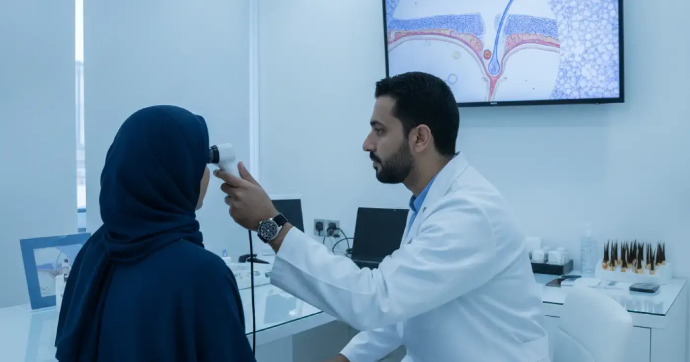
Learn how microscopic hair follicle analysis (trichoscopy) helps diagnose hair loss causes, identify scalp conditions, and guide targeted treatments.
Read article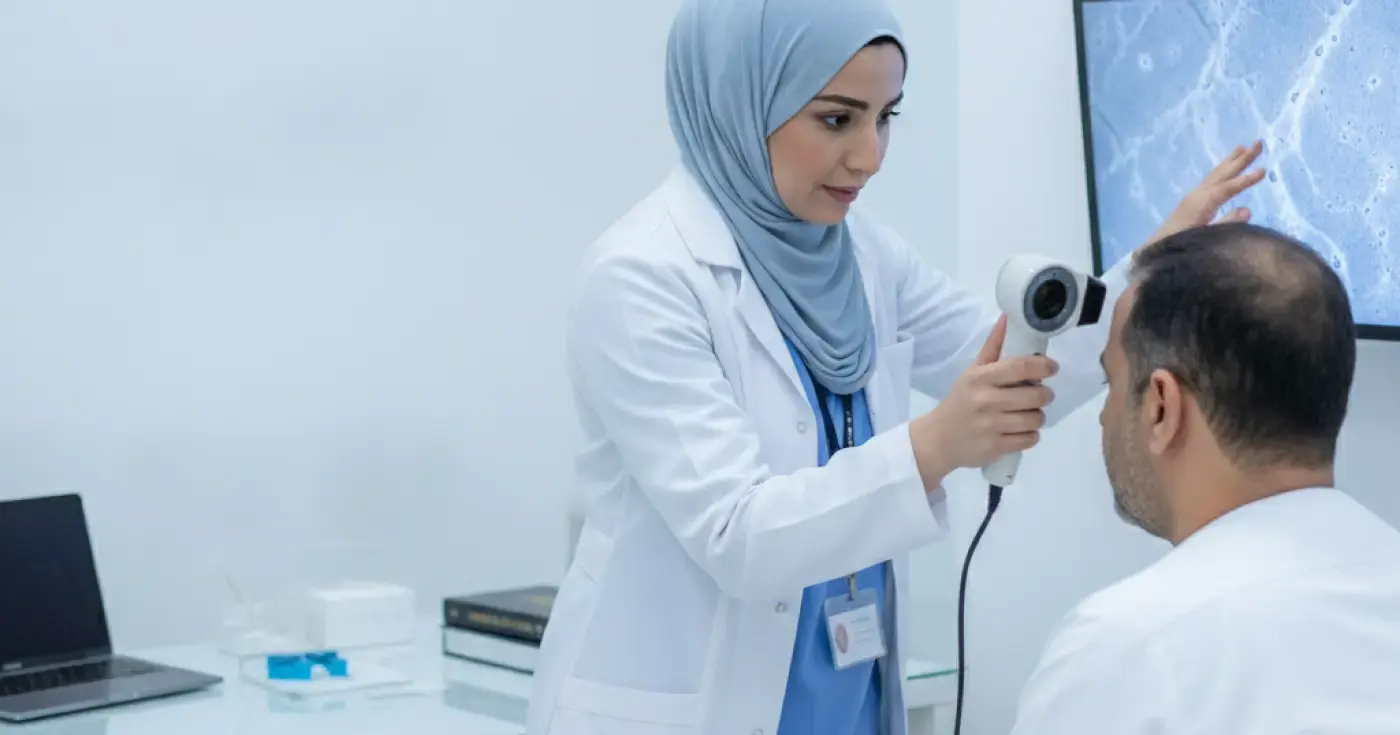
Learn how trichoscopy (microscopic scalp analysis) helps dermatologists accurately differentiate alopecia areata from other hair loss types.
Read article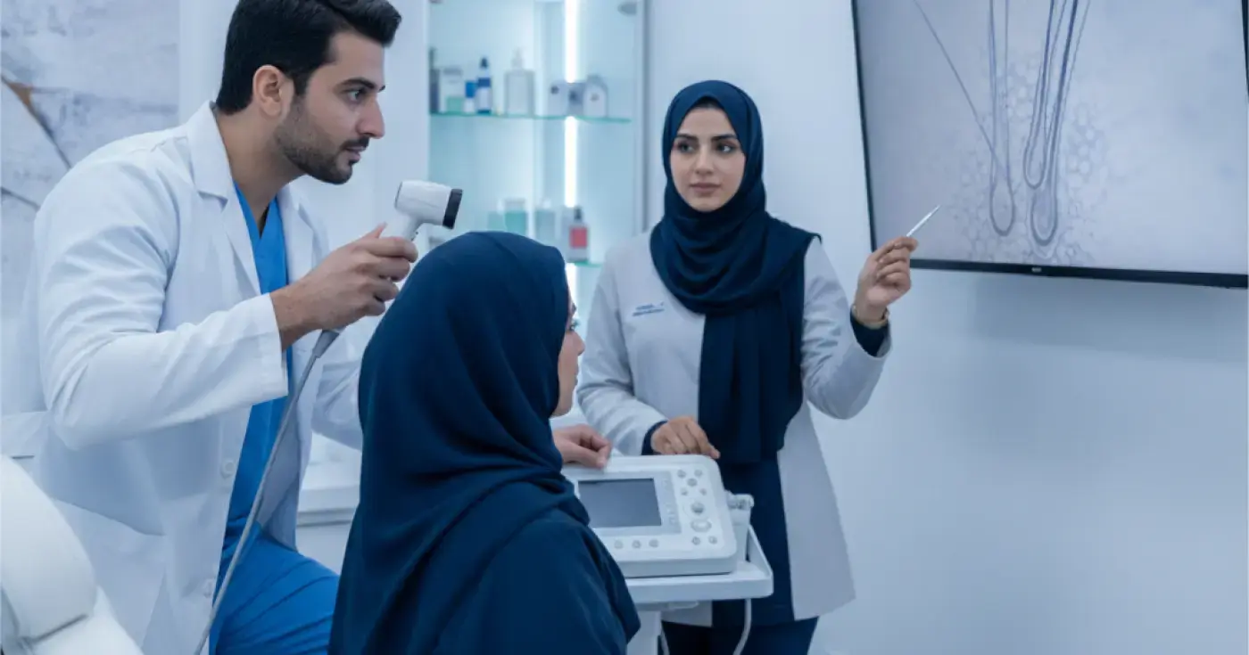
Discover how scalp trichoscopy (microscopic examination) helps dermatologists diagnose hair loss, alopecia, and scalp inflammation accurately and painlessly.
Read articleExplore the topic