
Lahalak Medical PlatformDifferentiate Care
Learn how microscopic hair follicle analysis (trichoscopy) helps diagnose hair loss causes, identify scalp conditions, and guide targeted treatments.
Advertisement
Hair loss and thinning are among the most common dermatological concerns, causing significant distress for both women and men. In the past, diagnosing the causes of hair loss relied heavily on simple visual inspection or a trial-and-error approach to treatment, which often led to delayed results and progression of the condition. Today, advanced dermatology has introduced a highly precise, non-invasive diagnostic tool known as trichoscopy, or microscopic hair follicle analysis. This painless procedure allows dermatologists to examine the scalp and hair follicles under high magnification, revealing vital clinical clues invisible to the naked eye and enabling highly customized, evidence-based treatment plans.
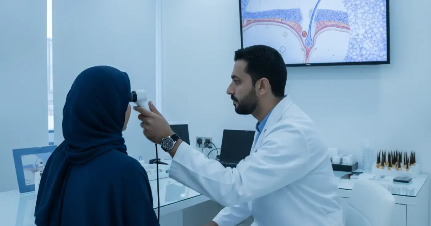
Trichoscopy is a non-invasive diagnostic technique that utilizes a specialized handheld device called a dermatoscope or a digital videodermatoscope. This technology magnifies the surface of the scalp, hair follicles, hair shafts, and surrounding blood vessels by 10x to over 70x. The primary goal of this analysis is to comprehensively evaluate scalp health, identify specific hair loss patterns, and differentiate between various types of alopecia, particularly distinguishing between scarring (cicatricial) alopecia, which causes permanent follicle destruction, and non-scarring alopecia, which is reversible with proper treatment.
The microscopic analysis works by projecting high-intensity polarized or non-polarized light onto the scalp through a magnifying lens connected to a high-definition digital camera. This allows the dermatologist to visualize both the superficial and deeper epidermal layers of the scalp without surgical intervention. The live images are transmitted directly to a computer screen, allowing both the physician and the patient to view the hair follicles clearly and document the findings for future comparison to monitor treatment progress.
Under high magnification, the dermatologist evaluates three main components: the hair shaft (its thickness, shape, and structural integrity), the follicular openings (whether they are open, empty, or fibrosed), and the perifollicular skin (looking for redness, scaling, or abnormal vascular patterns). These microscopic findings provide crucial diagnostic clues that guide the final clinical assessment.
During the microscopic analysis, the dermatologist looks for specific features known as "trichoscopic hallmarks." Each of these signs is associated with particular scalp conditions:
In a healthy scalp, hair shafts are relatively uniform in thickness. However, a significant variation in hair shaft diameter (where thick hairs sit next to very thin, miniaturized hairs) is the primary hallmark of androgenetic alopecia (pattern hair loss), indicating progressive follicular miniaturization driven by hormonal factors.
Yellow dots represent empty follicular infundibula filled with sebum and keratinous debris. They are highly characteristic of alopecia areata, indicating that the hair follicle is still present and viable but currently in an inactive (telogen) phase, suggesting a high potential for regrowth with targeted therapy.
Black dots are remnants of hair shafts that have broken off at the scalp surface due to acute inflammatory or autoimmune damage. Exclamation mark hairs are short, broken hairs that are thicker at the top and extremely thin near the scalp. Both are classic signs of active, progressive alopecia areata and indicate the need for prompt medical intervention.
These are short, tightly coiled hairs that result from mechanical pulling or incomplete hair breakage. They are characteristic findings in trichotillomania (hair-pulling disorder), helping clinicians differentiate it from autoimmune hair loss conditions.
The following table summarizes the key trichoscopic differences among the most common hair loss disorders:
| Condition | Hair Diameter Diversity | Yellow Dots | Black Dots / Exclamation Hairs | Perifollicular Scalp Condition |
|---|---|---|---|---|
| Androgenetic Alopecia | Very High (Miniaturization) | Rare (Only in advanced stages) | Absent | Normal, sometimes with increased sebum |
| Alopecia Areata | Low | Highly Common and Prominent | Present in active stages | Normal, non-scarring |
| Telogen Effluvium | Normal (Uniform thickness) | Absent | Absent | Completely normal and healthy |
| Scarring Alopecia | Absent (Loss of follicles) | Absent | Absent | Erythema, scaling, and loss of follicular openings |
Microscopic analysis is highly effective in diagnosing a wide range of hair and scalp disorders, including:
To ensure accurate results and clear visualization of the scalp, patients should follow these preparation guidelines:
Trichoscopy is a quick, comfortable, and completely painless office procedure. The typical steps include:
Historically, dermatologists had to rely on a scalp biopsy (removing a small skin sample under local anesthesia) to diagnose complex hair loss. While a biopsy remains necessary for certain scarring alopecias, trichoscopy offers a superior first-line alternative because it is completely non-invasive, painless, leaves no scars, provides immediate results, and allows for easy digital tracking of treatment efficacy over time.
You should consider scheduling a microscopic hair and scalp analysis if you experience:
Environmental factors in Saudi Arabia and the wider Gulf region—such as high temperatures, intense UV exposure, humidity, and the use of desalinated water—can impact scalp health. To protect your hair follicles, consider installing a shower filter to reduce chlorine and heavy metals, wear a hat or use an umbrella to shield your scalp from direct sunlight, maintain a balanced diet rich in proteins, iron, and zinc, and avoid tight hairstyles that cause traction alopecia.
Clear, direct answers
Advertisement
Continue exploring
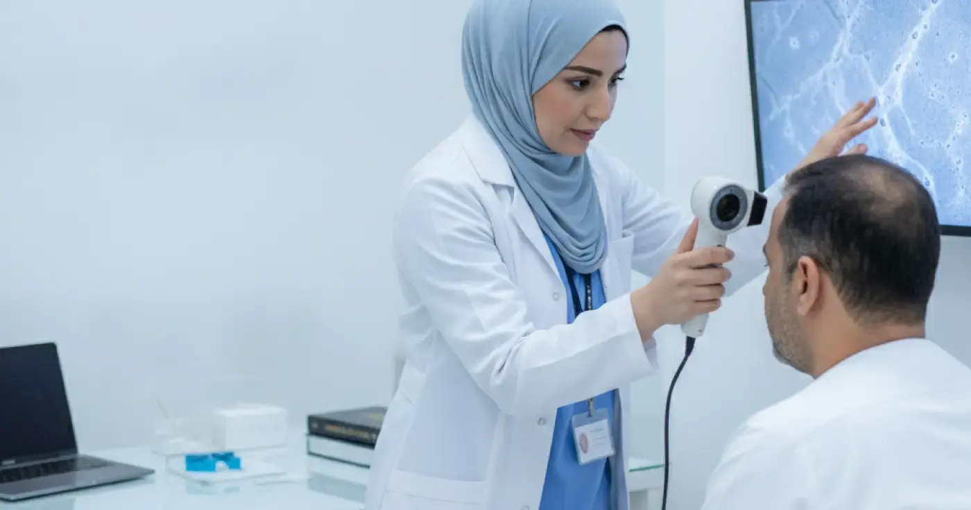
Learn how trichoscopy (microscopic scalp analysis) helps dermatologists accurately differentiate alopecia areata from other hair loss types.
Read article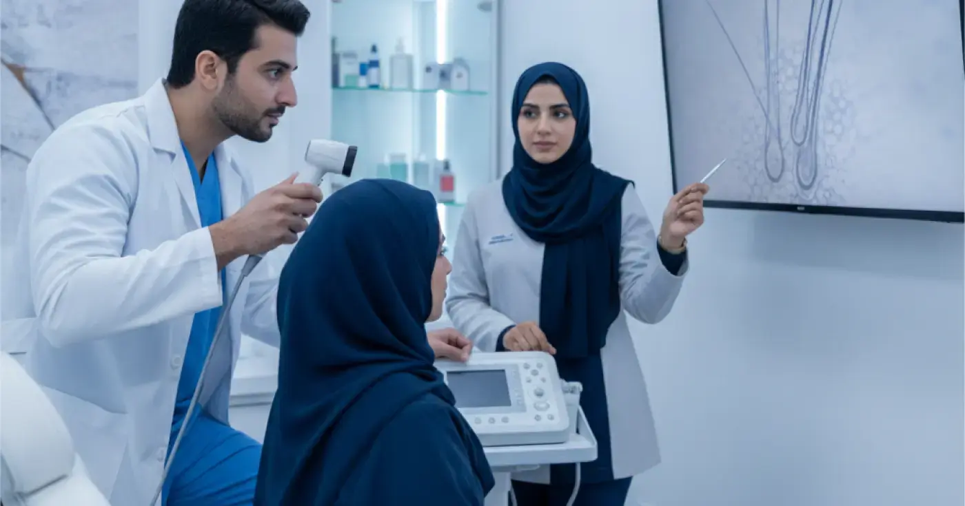
Discover how scalp trichoscopy (microscopic examination) helps dermatologists diagnose hair loss, alopecia, and scalp inflammation accurately and painlessly.
Read article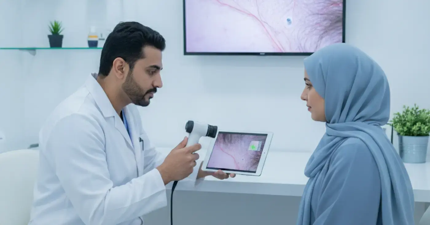
Learn how microscopic hair diagnosis (trichoscopy) helps dermatologists identify the root causes of hair loss and design effective treatment plans.
Read articleExplore the topic