
Lahalak Medical PlatformDifferentiate Care
Learn how microscopic hair diagnosis (trichoscopy) helps dermatologists identify the root causes of hair loss and design effective treatment plans.
Advertisement
Hair loss is a widespread concern affecting millions of men and women globally, often causing significant emotional distress. Historically, diagnosing the causes of hair loss relied heavily on basic visual inspection and clinical guesswork, which sometimes led to inaccurate diagnoses and ineffective treatments. With rapid advancements in dermatology, microscopic diagnosis of hair loss has emerged as a revolutionary tool, allowing specialists to see beyond the naked eye and identify precise underlying causes.
Understanding the root of the problem is the first and most crucial step toward successful recovery. Microscopic examination of the scalp and hair follicles helps differentiate between various types of hair loss—whether caused by genetics, autoimmune responses, physical stress, or nutritional deficiencies—paving the way for highly customized treatment plans.
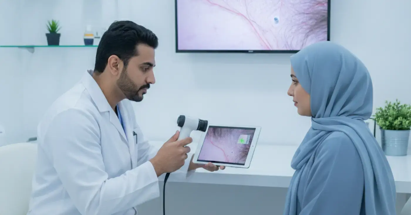
Microscopic hair diagnosis is a non-invasive medical technique that utilizes advanced optical magnification devices to examine the scalp, hair follicles, and hair shafts. This technology magnifies the target area from 20 to over 100 times, revealing intricate details about scalp health and hair structure.
The diagnosis generally involves two primary techniques:
Today, a trichoscope is the most widely used tool in dermatological clinics. It is a handheld video microscope connected to a computer screen, enabling the doctor to capture high-resolution images and videos of the scalp in real-time without plucking any hair. This exam evaluates hair density, shaft diameter, and the condition of the skin surrounding the follicles.
This is a semi-invasive method that involves gently plucking a small sample of hairs (usually 50 to 100) from different areas of the scalp to examine their roots under a light microscope. It helps determine the ratio of hair in various growth phases (anagen, catagen, and telogen), which is highly beneficial for diagnosing acute or chronic telogen effluvium.
The microscopic features of the scalp and hair vary significantly depending on the underlying cause of shedding. By analyzing these features, dermatologists can classify the condition accurately:
Under the microscope, androgenetic alopecia is characterized by significant variation in hair shaft diameter (thick hairs mixed with very thin, miniaturized ones), known as "follicular miniaturization." The doctor will also observe an increase in single-hair follicular units instead of the healthy, natural groupings of 2 to 4 hairs.
In this condition, microscopic analysis reveals a marked increase in the percentage of hair follicles in the resting (telogen) phase compared to the active growth phase. The scalp skin and individual hair shafts typically appear normal, without signs of severe inflammation or follicular scarring.
Alopecia areata presents with classic microscopic markers, such as "exclamation mark hairs"—short hairs that are narrow at the base and wider at the tip. Yellow and black dots are also commonly visible, representing empty follicular openings filled with sebum or remnants of broken hairs resulting from autoimmune attacks.
This is considered a dermatological emergency because it can lead to permanent hair loss. Microscopic examination shows a complete absence of follicular openings, replaced by fibrous scar tissue, often accompanied by active inflammation, redness, or scaling around the remaining follicles. Prompt medical intervention is vital to halt progression.
During the examination, the specialist focuses on identifying several vital markers to formulate a definitive diagnosis:
The following table outlines the key differences observed under the microscope to help differentiate between various hair loss conditions:
| Hair Loss Type | Follicular Unit Status | Hair Shaft Diameter Diversity | Key Microscopic Markers |
|---|---|---|---|
| Androgenetic Alopecia | Present, with increased single-hair units | Very High (Miniaturization) | Yellow dots, abundant vellus hairs |
| Alopecia Areata | Present, but empty in affected patches | Low | Exclamation mark hairs, black & yellow dots |
| Telogen Effluvium | Completely normal | Low to None | Increased ratio of telogen roots |
| Cicatricial Alopecia | Absent, replaced by fibrous tissue | Irregular | Loss of follicular ostia, erythema, scaling |
To ensure highly accurate results during your microscopic diagnosis, consider the following preparation steps before your dermatologist consultation:
Microscopic diagnosis offers numerous clinical benefits, making it the gold standard in modern hair clinics:
While shedding a certain number of hairs daily is normal, several warning signs warrant booking a professional microscopic evaluation:
Microscopic hair diagnosis represents a cornerstone of modern dermatology, transforming hair restoration from trial-and-error into a precise science. Early diagnosis is key to preserving your hair and restoring its vitality. Alongside professional clinical care, maintaining a balanced diet rich in essential vitamins, managing stress, and avoiding harsh chemical treatments will help protect your hair follicles and support long-term hair health.
Clear, direct answers
Advertisement
Continue exploring
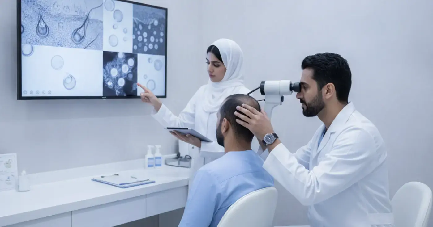
Learn how trichoscopy (microscopic hair analysis) helps dermatologists detect follicle weakness and identify the exact causes of hair loss non-invasively.
Read article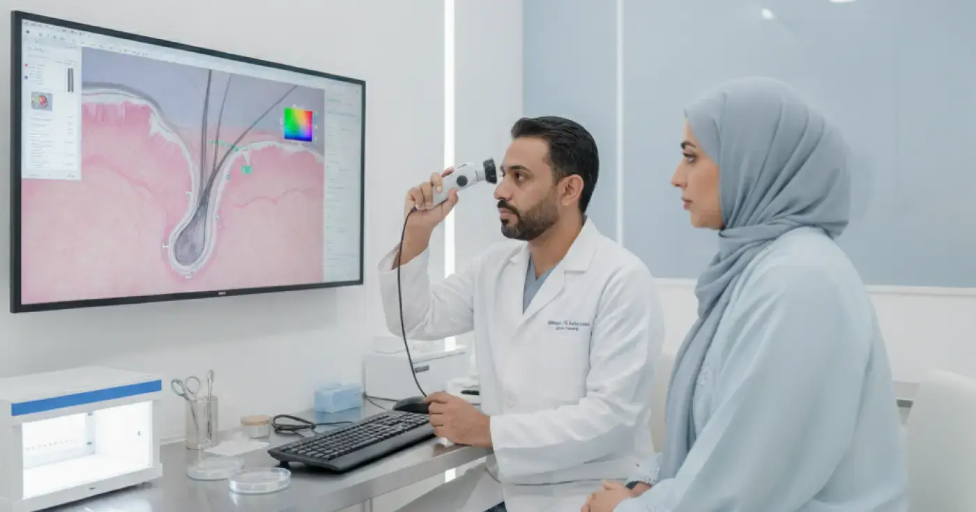
Discover how microscopic hair analysis (trichoscopy) helps dermatologists diagnose hair loss, alopecia, and scalp conditions accurately.
Read article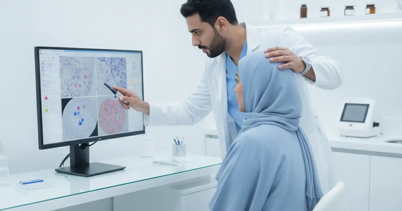
Discover how microscopic hair analysis (Trichoscopy) diagnoses hair loss causes accurately to guide effective, personalized treatment.
Read articleExplore the topic
The Role of Trichoscopy in Detecting Hair Follicle Weakness and Growth IssuesElixir · Health PanoramaMicroscopic Hair Analysis: A Comprehensive Guide to Diagnosing Scalp and Hair DisordersElixir · Health PanoramaMicroscopic Diagnosis of Hair Problems: How Trichoscopy Reveals Hair Loss CausesElixir · Health Panorama