
Lahalak Medical PlatformDifferentiate Care
Discover how scalp trichoscopy (microscopic examination) helps dermatologists diagnose hair loss, alopecia, and scalp inflammation accurately and painlessly.
Advertisement
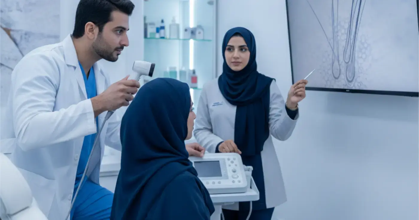
Scalp trichoscopy, medically known as trichoscopy, is one of the most prominent modern, non-invasive diagnostic techniques in dermatology. This technique relies on using a digital dermoscope to magnify the scalp and hair follicles by tens to hundreds of times, allowing the dermatologist to see fine details that cannot be seen with the naked eye.
In the past, diagnosing hair loss problems relied heavily on simple clinical examination or resorting to more painful options such as a scalp biopsy. Today, microscopic examination has revolutionized the way hair and scalp health is evaluated, making it easier to develop an accurate and customized treatment plan for each case.
The examination relies on projecting polarized or non-polarized light through a magnifying lens connected to a high-resolution camera. The physician gently places the scope on different areas of the scalp, and the images are transmitted directly to a computer screen. This allows for precise analysis of the following factors:
Microscopic examination helps diagnose and differentiate a wide range of hair and scalp disorders, including:
Pattern hair loss appears microscopically as a high diversity in hair shaft diameter (the presence of thick hairs alongside very thin, short hairs), as well as an increase in the number of follicles containing only a single hair instead of normal hair groups.
Alopecia areata is characterized by highly distinctive microscopic signs such as "exclamation mark hairs" (short hairs that are narrower at the base and thicker at the top), as well as black dots and yellow dots representing empty hair follicles.
Scarring alopecia is a serious condition that requires rapid diagnosis to prevent permanent hair loss. Microscopic examination in this case shows a complete absence of follicular openings with skin fibrosis, redness, and scaling around the remaining follicles.
Trichoscopy makes it easy to see yellow or white greasy scales adhering to the scalp and to determine the severity of the associated skin inflammation.
Psoriasis appears microscopically as thick, dry, silvery scales, with very characteristic patterns of dilated and tortuous blood vessels under the skin.
The following table illustrates the fundamental differences between relying on traditional clinical examination and modern microscopic examination of the scalp:
| Feature | Traditional Clinical Exam | Scalp Trichoscopy |
|---|---|---|
| Diagnostic Accuracy | Limited and depends on visible symptoms only | Very high, revealing invisible microscopic changes |
| Need for Biopsy | Higher frequency to confirm diagnosis | Significantly reduces the need for surgical biopsy |
| Treatment Monitoring | Relies on approximate visual assessment | Precise measurement of hair thickness and density in mm |
| Early Detection | Detects the problem after significant hair loss | Detects follicular weakening before actual hair loss |
During the hair follicle analysis under the microscope, the dermatologist focuses on identifying specific signs that help formulate the final diagnosis:
The microscopic examination session is comfortable, quick, and completely painless. The steps are as follows:
The patient is advised not to wash their hair on the day of the examination to avoid removing scales or natural sebum secretions that may help in diagnosis. Avoiding styling products or temporary dyes is also recommended.
The patient sits comfortably, and the doctor parts the hair into sections, gently passing the dermoscope lens over the scalp. High-resolution photos are taken of multiple areas (frontal, parietal, and occipital) for comparison.
The doctor analyzes the images and shares them with the patient on the screen to explain the nature of the condition, and then establishes the appropriate hair loss treatment plan based on the visual results.
Microscopic examination is 100% safe, as it does not involve any skin penetration or the use of harmful radiation. It is suitable for all ages, including children and pregnant women. However, it should be noted that in some complex or rare cases of scarring alopecia, microscopic examination alone may not be sufficient, forcing the treating physician to request a small skin biopsy to confirm the histological diagnosis accurately.
To ensure the most accurate results from your scalp trichoscopy, it is recommended to follow these guidelines:
Scalp trichoscopy represents an indispensable diagnostic tool in modern dermatology clinics. This examination allows a transition from visual guesswork to precise, evidence-based scientific diagnosis. If you suffer from unexplained hair loss, persistent itching, or the appearance of scales and inflammation on your scalp, consulting a specialist dermatologist and undergoing a microscopic examination is the first and correct step toward restoring your hair health and self-confidence.
Clear, direct answers
Advertisement
Continue exploring
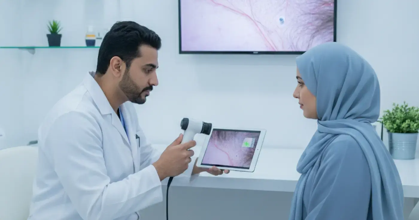
Learn how microscopic hair diagnosis (trichoscopy) helps dermatologists identify the root causes of hair loss and design effective treatment plans.
Read article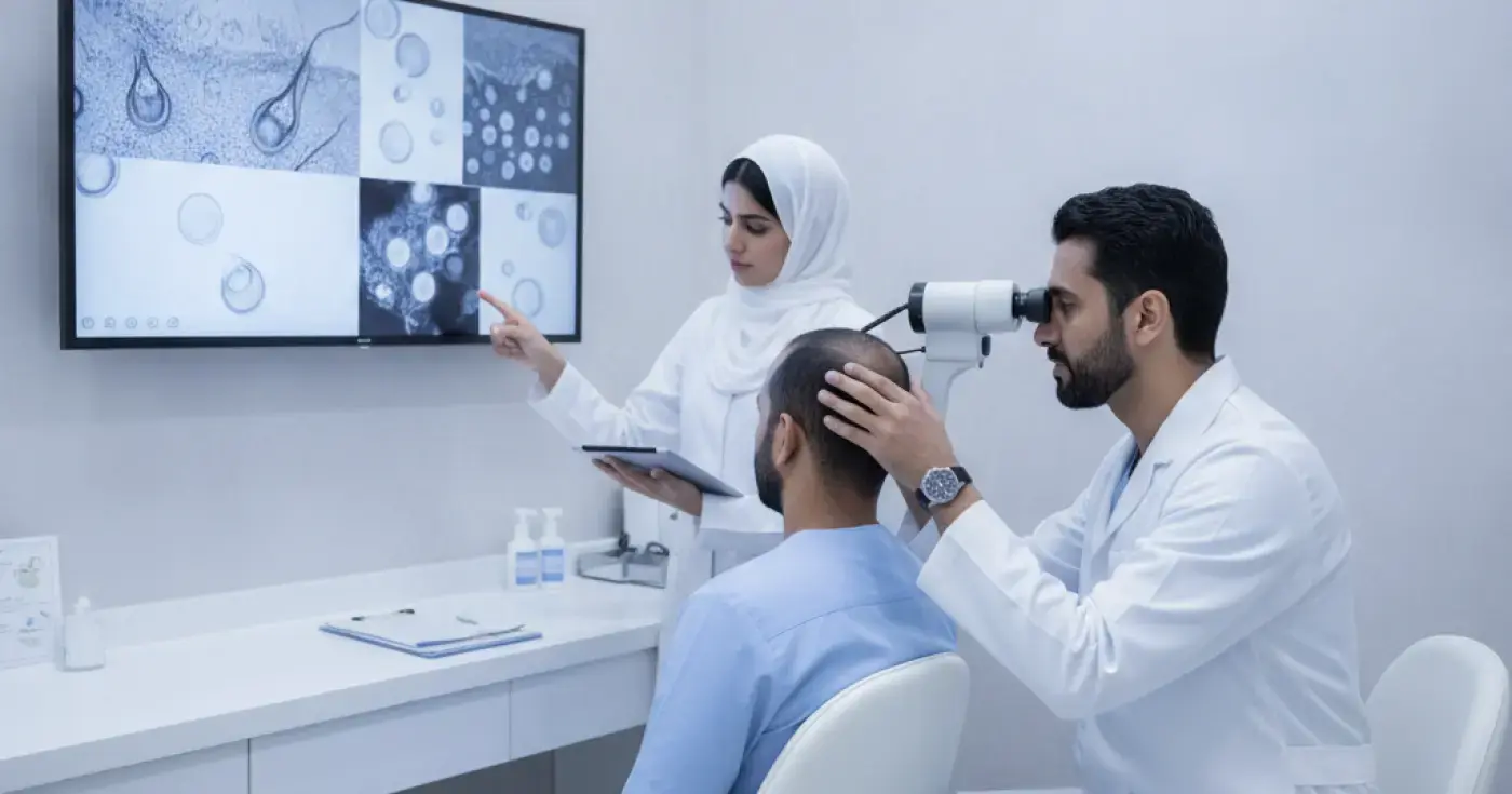
Learn how trichoscopy (microscopic hair analysis) helps dermatologists detect follicle weakness and identify the exact causes of hair loss non-invasively.
Read article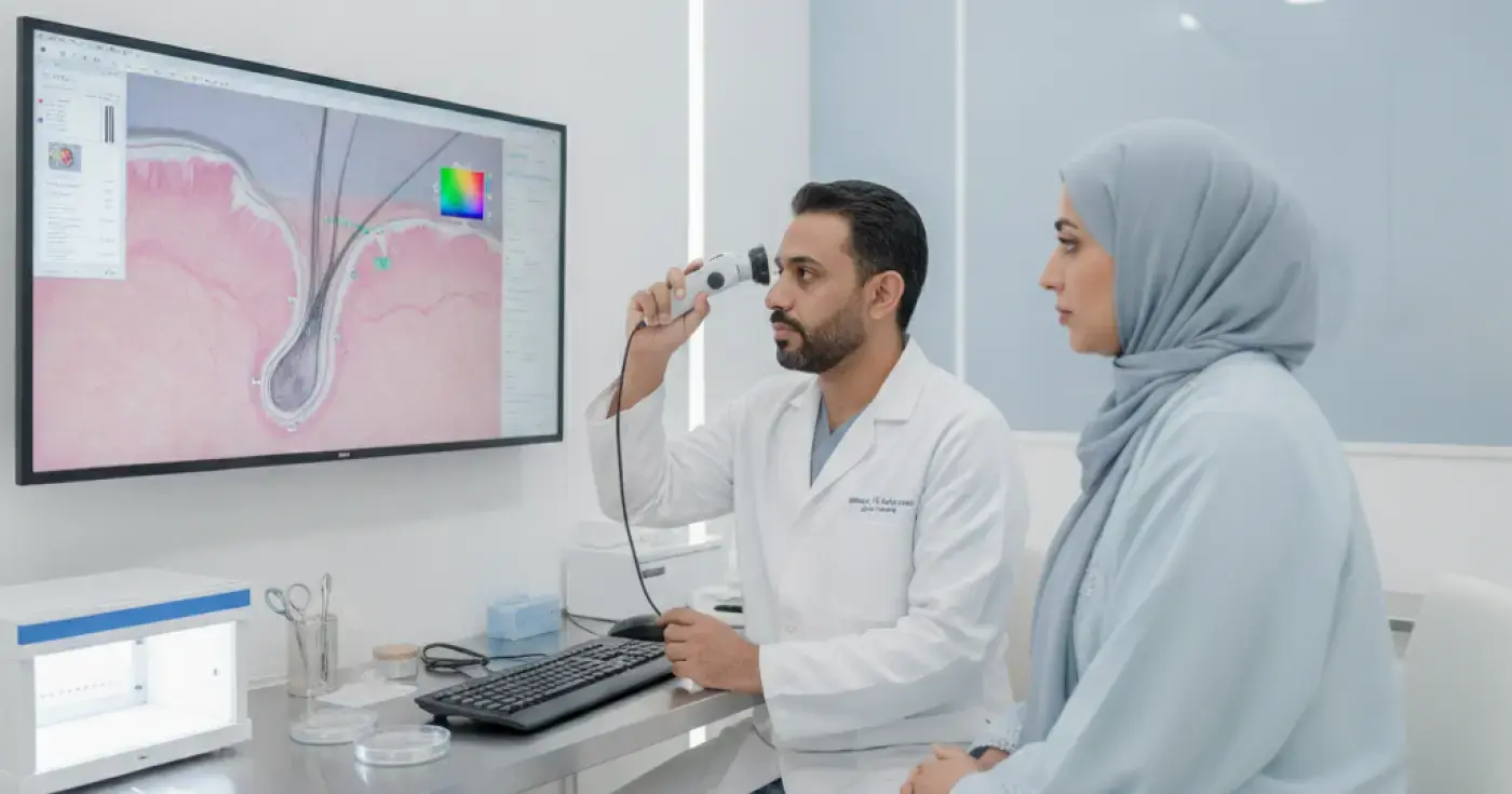
Discover how microscopic hair analysis (trichoscopy) helps dermatologists diagnose hair loss, alopecia, and scalp conditions accurately.
Read articleExplore the topic
Microscopic Diagnosis of Hair Loss: How Doctors Pinpoint the CauseElixir · Health PanoramaThe Role of Trichoscopy in Detecting Hair Follicle Weakness and Growth IssuesElixir · Health PanoramaMicroscopic Hair Analysis: A Comprehensive Guide to Diagnosing Scalp and Hair DisordersElixir · Health Panorama