
Lahalak Medical PlatformDifferentiate Care
A complete medical guide on the clinical steps, biopsy types, pathology results, and treatment options after detecting an abnormal mole.
Advertisement
Moles (nevi) are common skin growths that most people have, and the vast majority are benign and harmless. However, during routine skin checks or self-examinations, a dermatologist may identify an "abnormal" or atypical mole. Discovering an abnormal mole does not automatically mean you have skin cancer; rather, it is a critical preventive step that allows medical professionals to intervene early and prevent future complications.
In Saudi Arabia, public health initiatives place a strong emphasis on skin health and sun protection due to the high levels of UV radiation. Understanding the clinical pathway that follows the detection of an atypical mole helps patients navigate their care with confidence and peace of mind.
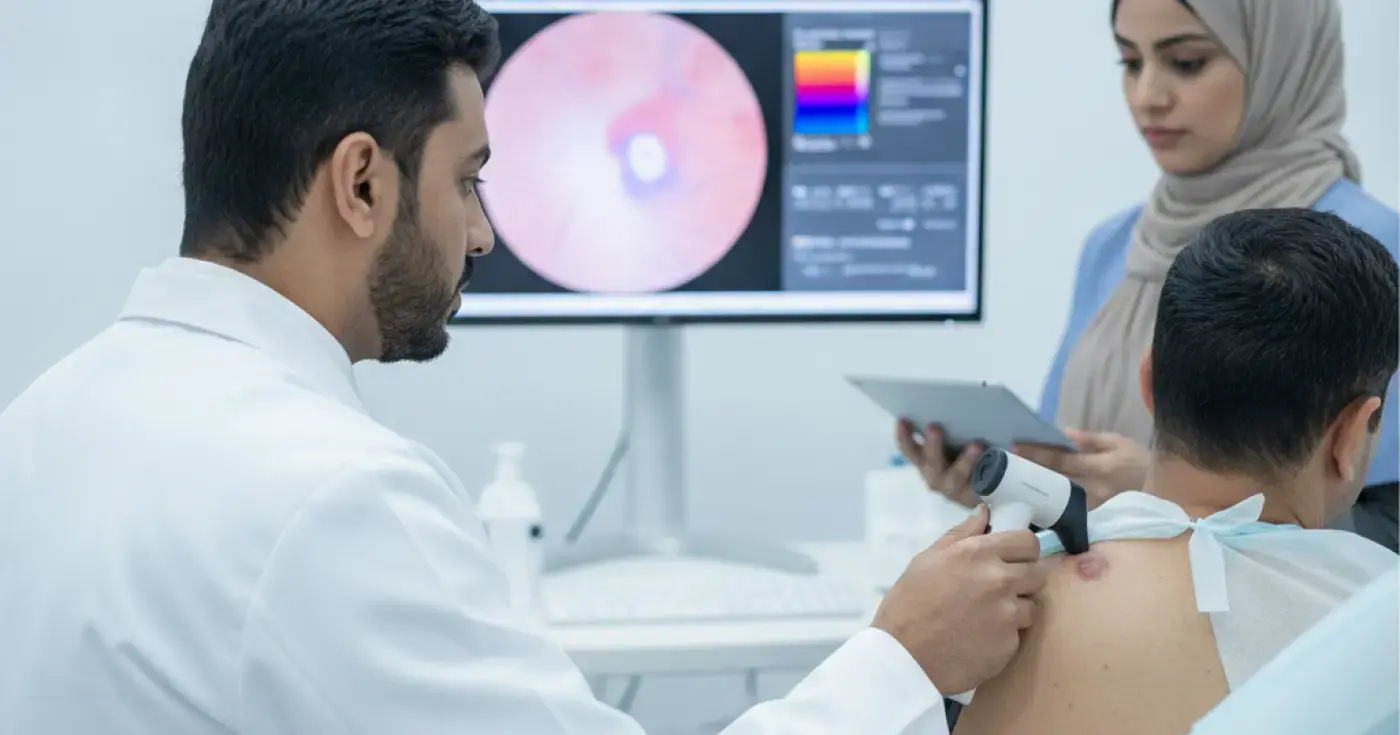
When a dermatologist identifies a suspicious mole, they do not rely solely on the naked eye. The first step is a detailed evaluation using a specialized tool called a dermatoscope. This non-invasive procedure, known as fحص الشامات بالمنظار (Dermoscopy), uses polarized light and high-magnification lenses to visualize pigment patterns and blood vessels beneath the skin's surface.
During this examination, the physician evaluates the mole using the established "ABCDE" criteria:
If the dermoscopy reveals atypical features, or if a mole has changed significantly over time, the standard medical recommendation is to perform a خزعة الجلد (Skin Biopsy). A biopsy is the only definitive way to diagnose the nature of a mole; visual inspection alone cannot provide a final diagnosis.
The biopsy involves removing a portion or the entirety of the mole under local anesthesia so that the tissue can be analyzed under a microscope. It is a quick, safe, and outpatient procedure performed in the clinic.
The choice of biopsy technique depends on the mole's size, location, and the level of clinical suspicion. The table below outlines the primary biopsy methods:
| Biopsy Type | Procedure Description | Common Indications | Expected Recovery Time |
|---|---|---|---|
| Shave Biopsy | The doctor uses a thin blade to shave off the superficial layers of the skin. | Raised moles with low suspicion of deep invasion. | 7 to 10 days (no stitches required). |
| Punch Biopsy | A circular tool is used to remove a deeper, cylindrical core of skin tissue. | Smaller moles where full-thickness evaluation is needed. | 10 to 14 days (may require 1-2 stitches). |
| Excisional Biopsy | The entire mole is removed along with a small margin of healthy skin using a scalpel. | Highly suspicious lesions or suspected melanoma. | 2 to 3 weeks (requires surgical sutures). |
Once the tissue sample is collected, it is preserved in formalin and sent to a pathology laboratory. A dermatopathologist—a pathologist specializing in skin diseases—processes the tissue, cuts it into microscopic slices, stains it, and examines it under a high-power microscope.
The pathologist evaluates the cellular structure, the arrangement of melanocytes (pigment-producing cells), and whether there are signs of abnormal cell division. This highly precise process typically takes 3 to 7 business days, and the resulting pathology report guides all subsequent clinical decisions.
The pathology report will classify the mole into one of several categories:
Based on the pathology findings, the dermatologist will establish a personalized management plan:
For moderately to severely dysplastic nevi or early-stage melanoma, a wider surgical excision (استئصال الشامات) is performed. This involves removing the biopsy site along with a predetermined margin of healthy surrounding skin to ensure no abnormal cells remain, significantly reducing the risk of recurrence.
In advanced cases, patients are referred to a multidisciplinary oncology team to evaluate regional lymph nodes and discuss advanced therapeutic options, such as immunotherapy or targeted therapies, available at specialized medical centers in the Kingdom.
Proper wound care is essential to promote healing, minimize scarring, and prevent infection. Key recommendations include:
Having an atypical mole should encourage a proactive approach to skin health. Performing a monthly الفحص الذاتي للشامات (Self-examination of moles) is a highly effective way to spot new or changing lesions early. Use a well-lit room and mirrors to check all areas of your body, including your back and scalp.
In addition to self-checks, schedule an annual professional skin exam with your dermatologist, especially if you have a family history of skin cancer, fair skin, or a large number of moles.
Clear, direct answers
Advertisement
Continue exploring
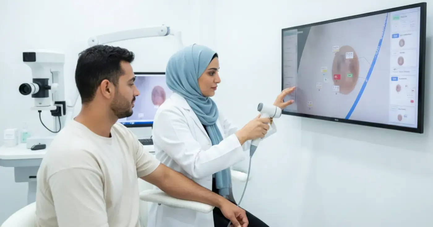
Discover the clinical importance of mole screening for individuals with multiple moles. Learn about the ABCDE rule, dermoscopy, and digital mole mapping.
Read article
Learn how often to get your moles checked, the ABCDE rule for self-exams, and when to see a dermatologist to prevent skin cancer.
Read article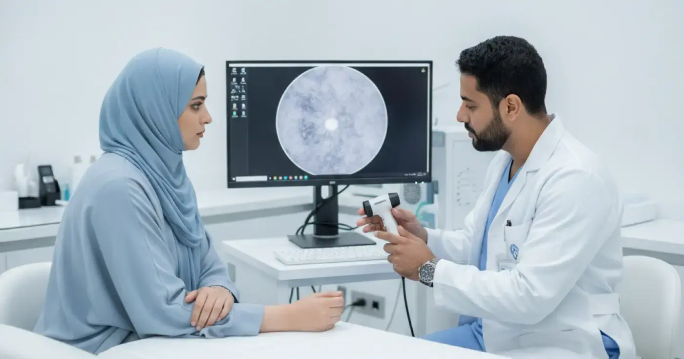
Learn about dermoscopy for mole examination, how it works, what to expect during your clinic visit, and its role in early skin cancer detection.
Read articleExplore the topic