
Lahalak Medical PlatformDifferentiate Care
Learn about dermoscopy for mole examination, how it works, what to expect during your clinic visit, and its role in early skin cancer detection.
Advertisement
Moles (nevi) are common skin growths that most people develop over time. While the vast majority of moles are completely benign and harmless, some can undergo subtle changes that may indicate the development of atypical cells or skin cancers, such as melanoma. This is where dermoscopy mole examination plays a pivotal, non-invasive diagnostic role.
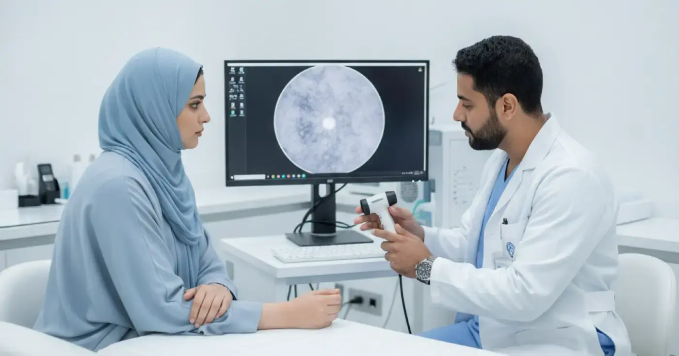
A dermatoscope is a specialized handheld medical device equipped with high-quality magnifying lenses and a polarized lighting system. This tool allows dermatologists to inspect the outer and immediate sub-surface layers of the skin with extreme precision, revealing pigment patterns and vascular structures invisible to the naked eye. This technology is essential for the early detection of skin cancer and helps prevent unnecessary surgical biopsies.
The primary goal of regular mole screening is the early detection of malignant skin tumors, particularly melanoma. Melanoma is one of the most aggressive forms of skin cancer, yet it is highly curable when identified and treated in its earliest stages. Dermoscopy helps in several ways:
A dermatoscope relies on advanced optical technology combining magnification (typically ranging from 10x to over 100x) with specialized lighting. There are two main types of illumination used in modern devices:
1. Polarized Light: This allows the specialist to visualize deeper skin structures without needing to apply a liquid or gel to the skin, as polarized light directly penetrates the stratum corneum.
2. Non-polarized Light: This requires a contact liquid (such as medical alcohol, oil, or gel) on the skin to reduce surface reflection, facilitating a clear view of the superficial layers of the epidermis.
Through these mechanisms, the dermatologist can evaluate pigment symmetry, pigment network distribution, irregular dots or globules, and vascular structures, which are critical details that cannot be seen with the naked eye.
While routine skin checks are beneficial for everyone, certain individuals have a higher risk of developing atypical moles or skin cancer and should be monitored closely by a dermatologist. Risk factors include:
A dermoscopy examination is a simple, quick, and entirely painless procedure. Here is what you can expect before, during, and after your appointment:
No complex preparation is required. However, patients are advised to avoid wearing makeup or nail polish (if moles are present under the nails or on the face) to ensure clear visibility. Wearing easily removable clothing is also recommended for a full-body skin check.
The dermatologist examines the moles one by one using the handheld dermatoscope. If a digital system is used, the device is connected to a computer screen to display and record magnified images of the moles. A full-body check typically takes between 15 to 30 minutes, depending on the number of moles.
The doctor discusses the findings immediately. If all moles are normal, a routine follow-up is scheduled (usually in 12 months). If a suspicious or atypical mole is detected, the doctor may recommend mole removal for a biopsy to rule out malignancy.
The table below outlines the key differences between a standard naked-eye visual exam and a specialized dermoscopy examination:
| Feature | Traditional Visual Exam (Naked Eye) | Dermoscopy Examination (Dermatoscope) |
|---|---|---|
| Magnification Level | None (1x) | High (10x to over 100x) |
| Deep Structure Visibility | Limited to the outer skin surface | Penetrates the outer layer to view deep structures and vessels |
| Early Detection Accuracy | Moderate; may miss very early cellular changes | Very high; detects cellular changes before they surface | May be higher due to general visual uncertainty | Significantly lower, as it accurately rules out benign moles |
| Digital Tracking | Difficult and unreliable for future comparison | Highly reliable through digital image archiving and comparison |
Self-monitoring is an essential preventive step that complements professional clinical exams. Dermatologists recommend using the international "ABCDE" rule to check your moles monthly:
Dermatology has experienced a significant technological shift with the integration of Total Body Mapping and digital dermoscopy. These systems capture high-resolution photos of the patient's entire body, mapping the exact location of every mole. In subsequent visits, automated software compares the images to flag any new or changing lesions. Artificial intelligence (AI) algorithms are also beginning to assist clinicians by analyzing pigment patterns, further enhancing diagnostic accuracy.
Prevention remains the most effective defense against skin cancer and abnormal mole changes. Follow these dermatologist-approved guidelines:
Remember, a professional dermoscopy exam is a quick, safe, and potentially life-saving preventive measure. If you notice any changing or suspicious moles, consult a qualified dermatologist for a professional evaluation.
Clear, direct answers
Advertisement
Continue exploring
Learn the warning signs of changing moles. Discover the ABCDE criteria, normal vs. abnormal changes, and when to see a dermatologist.
Read article
Learn how to tell the difference between benign and suspicious moles using the ABCDE rule. Discover warning signs, prevention tips, and when to see a doctor.
Read article
Learn the warning signs in moles using the ABCDE rule. Discover when to consult a dermatologist for skin cancer screening and evaluation.
Read article