
Lahalak Medical PlatformDifferentiate Care
Learn how microscopic hair analysis (trichoscopy) accurately diagnoses hair loss causes and guides dermatologists to select the best treatment.
Advertisement
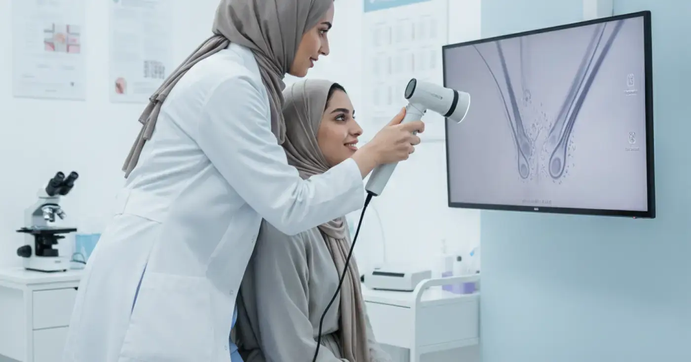
Hair loss and scalp disorders are among the most common concerns addressed in dermatology clinics. In the past, diagnosing these conditions relied heavily on visual inspection and simple hair pull tests, which often lacked the precision needed to identify the underlying cause. With technological advancements, microscopic hair analysis (trichoscopy) has emerged as a revolutionary, non-invasive diagnostic tool that allows dermatologists to examine hair follicles and the scalp with extreme precision.
Trichoscopy utilizes a specialized handheld device called a dermatoscope or a digital trichoscope. This device magnifies the scalp and hair structures from 10x to over 100x magnification. By projecting these high-resolution images onto a monitor, dermatologists can analyze the hair shaft, follicular openings, the surrounding skin, and the microvascular patterns of the scalp. This detailed view reveals subtle pathological changes that are completely invisible to the naked eye.
Microscopic hair analysis is highly effective in differentiating between various types of hair loss, which is crucial because their treatments differ significantly. Key conditions diagnosed include:
Compared to traditional diagnostic methods, trichoscopy offers numerous benefits for both patients and clinicians:
During the examination, the specialist focuses on several key anatomical features:
The procedure is straightforward and comfortable:
| Feature / Condition | Androgenetic Alopecia | Alopecia Areata | Telogen Effluvium |
|---|---|---|---|
| Hair Diameter | Highly variable (miniaturization) | Uniformly thin or broken | Mostly uniform |
| Key Microscopic Signs | Brown peripilar sign, yellow dots | Exclamation mark hairs, black dots | Empty hair follicles, upright regrowing hairs |
| Follicular Openings | Preserved | Preserved (often filled with sebum/debris) | Preserved |
| Inflammatory Signs | Minimal to none | Mild perifollicular inflammation | None |
Accurate diagnosis is the cornerstone of successful treatment. For instance, if trichoscopy reveals early signs of androgenetic alopecia, the doctor may prescribe topical minoxidil or oral DHT blockers. If active alopecia areata is detected, localized corticosteroid injections may be recommended. Furthermore, for patients undergoing therapies like PRP therapy for hair, follow-up trichoscopy sessions provide visual proof of increased hair density and thickness, ensuring the treatment is working effectively.
It is highly recommended to seek a professional evaluation if you experience sudden or excessive hair shedding, localized bald patches, persistent scalp itching, scaling, or redness. Early intervention guided by microscopic diagnosis can prevent irreversible follicle damage, especially in scarring alopecia conditions.
Microscopic hair analysis has transformed the field of trichology, turning subjective assessments into an objective, science-based discipline. By pinpointing the exact nature of your hair or scalp condition, this advanced technology ensures you receive a personalized, highly effective treatment plan, saving time and preserving your hair health.
Clear, direct answers
Advertisement
Continue exploring
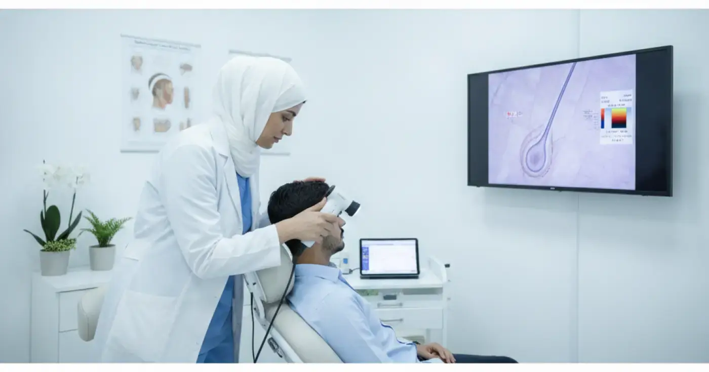
Discover the importance of microscopic hair and scalp analysis (trichoscopy) to diagnose hair loss causes and find the right treatment with dermatologists.
Read article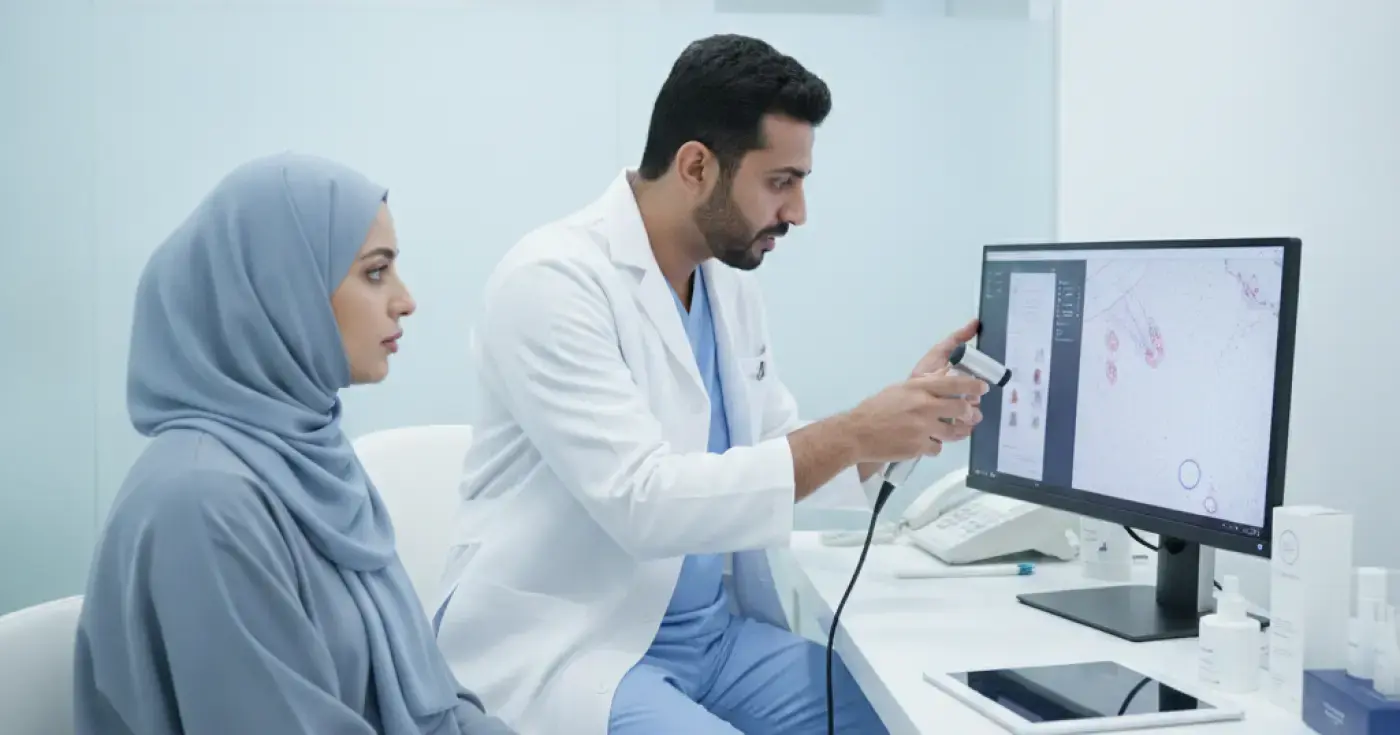
Discover how trichoscopy helps dermatologists diagnose dandruff, seborrheic dermatitis, and scalp inflammation for targeted, effective treatment.
Read article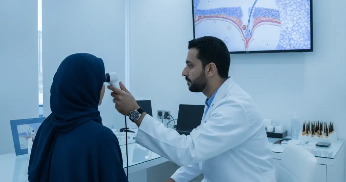
Learn how microscopic hair follicle analysis (trichoscopy) helps diagnose hair loss causes, identify scalp conditions, and guide targeted treatments.
Read articleExplore the topic