
Lahalak Medical PlatformDifferentiate Care
Explore safe medical methods for removing benign skin tumors, including cryotherapy, laser, and surgical excision, with expert recovery tips.
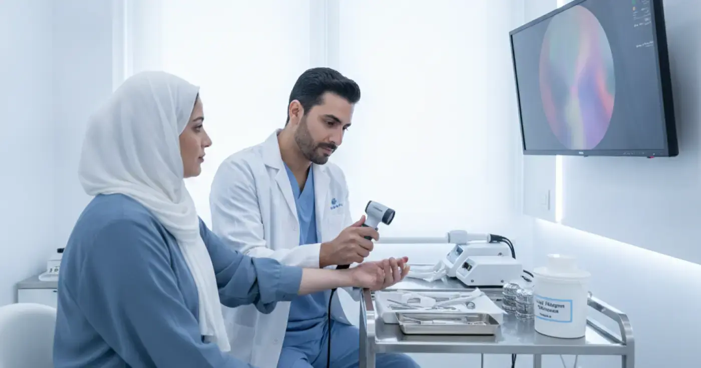
Advertisement
Benign skin tumors are highly common conditions encountered daily by dermatologists. These non-cancerous skin growths develop on or just beneath the skin surface. Although they do not pose an immediate threat to health, many patients choose to have them removed for cosmetic reasons, due to functional irritation caused by friction with clothing or jewelry, or to achieve absolute peace of mind through definitive histological confirmation.
Before exploring removal techniques, it is essential to identify the most common types of benign skin lesions treated in clinical settings:
The decision to remove a benign skin lesion is made collaboratively between the dermatologist and the patient, based on several key factors:
Dermatology clinics utilize several advanced techniques to remove benign skin tumors. The choice of method depends on the tumor's type, size, depth, and anatomical location:
This traditional and highly precise method is ideal for deep moles, lipomas, and larger lesions. Under local anesthesia, the dermatologist uses a scalpel to excise the entire tumor along with a narrow margin of healthy skin. The wound is then closed with fine cosmetic sutures. A major advantage of this technique is that it preserves the entire specimen for laboratory analysis.
Cryotherapy involves applying extreme cold, typically using liquid nitrogen (at approximately -196°C), directly to the skin lesion. This rapid freezing destroys the targeted cells, causing a small blister to form. The lesion eventually sloughs off naturally within one to two weeks. This method is highly effective for seborrheic keratoses and common warts.
This technique utilizes high-frequency electrical currents to burn and dehydrate the targeted tissue while simultaneously sealing blood vessels to prevent bleeding. Following electrosurgery, the doctor may use a spoon-shaped instrument called a curette to scrape away the treated tissue. It is highly suitable for skin tags and superficial lesions.
Laser removal is an advanced cosmetic option preferred for superficial lesions and small moles, particularly on the face. Utilizing precise lasers (such as the CO2 laser), the dermatologist vaporizes the tumor cells with minimal damage to the surrounding healthy tissue, significantly reducing scarring risk and accelerating healing.
RF ablation uses high-frequency radio waves to cut or coagulate tissue with extreme precision. It causes minimal bleeding, promotes rapid healing, and is ideal for removing protruding moles and skin tags on the face due to its excellent cosmetic outcomes.
The following table provides a quick comparison of the clinical techniques to help patients understand the key differences:
| Removal Method | Best Suited For | Anesthesia Type | Expected Recovery Time | Scarring Risk |
|---|---|---|---|---|
| Surgical Excision | Deep moles, lipomas, cysts | Local injection | 7 - 14 days (requires suture removal) | Moderate (fine linear scar) |
| Cryotherapy | Seborrheic keratosis, warts | Usually none | 7 - 10 days | Very low (temporary pigment change possible) |
| Electrosurgery | Skin tags, superficial lesions | Local cream or injection | 5 - 10 days | Low to Moderate |
| Laser Therapy | Superficial moles, facial lesions | Local cream or injection | 5 - 7 days | Very low |
| Radiofrequency | Protruding moles, skin tags | Local cream or injection | 5 - 7 days | Very low |
The process begins with a thorough consultation where the dermatologist evaluates the lesion, often using a dermatoscope. Patients must inform their doctor of any medical history, bleeding disorders, allergies to local anesthetics, or a tendency to develop keloid scars.
Most removals are performed as quick, outpatient procedures lasting between 15 and 30 minutes. The target area is thoroughly sanitized, local anesthesia is administered (via injection or topical cream), and the chosen technique is executed precisely to ensure a pain-free experience.
The quality of the final cosmetic outcome depends heavily on proper wound care. Key instructions include:
While these clinical procedures are exceptionally safe when performed by a qualified dermatologist, minor risks include:
Dermatologists adhere to a fundamental clinical rule: "Any excised tissue should be sent for laboratory analysis." Even when a lesion appears highly benign, a histopathological examination under a microscope is the only definitive way to confirm cell safety, providing absolute peace of mind and accurate medical documentation.
Removing benign skin tumors in a clinical setting is a safe, straightforward process that enhances comfort, appearance, and self-confidence. At LahaLak Medical Platform, we strongly advise against attempting to remove skin tags or moles at home using DIY kits, threads, or acidic home remedies. These unsafe practices often lead to severe infections, permanent scarring, or delayed diagnosis of serious conditions. Always consult a board-certified dermatologist to evaluate your skin and select the safest, most effective treatment.
Clear, direct answers
Advertisement
Continue exploring
Learn if benign skin lesions can become cancerous, discover the ABCDE warning signs, and understand when to consult a dermatologist.
Read article
Learn about benign skin lumps, common types like lipomas and cysts, and the warning signs that require an immediate visit to a dermatologist.
Read article
Learn about subcutaneous lipomas (benign fatty lumps), their causes, how they are diagnosed, and the latest surgical and non-surgical removal options.
Read article