Lahalak Medical PlatformDifferentiate Care
Discover signs of scalp ringworm in children, how it differs from alopecia and dandruff, and the role of fungal microscopic testing and culture.
Advertisement
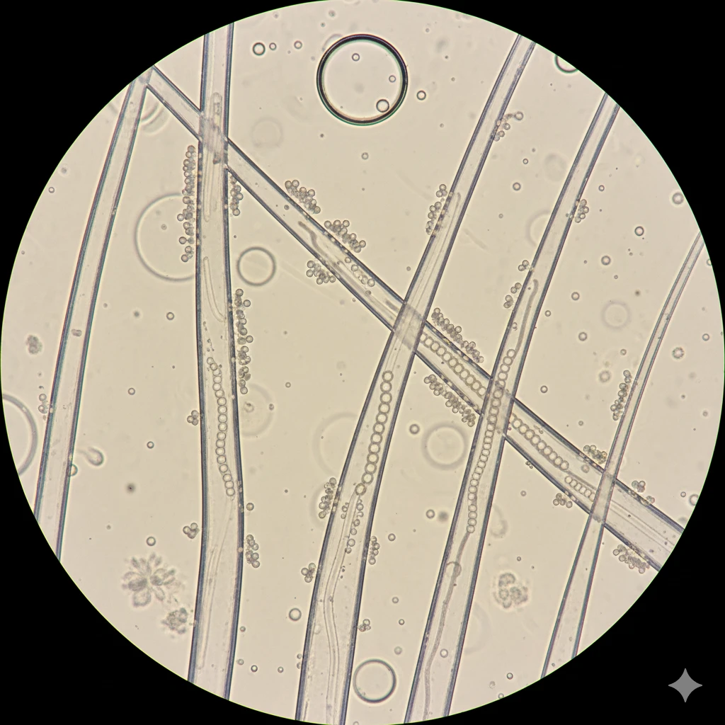
Tinea capitis, commonly known as scalp ringworm, is a frequent fungal infection affecting the scalp and hair shafts in children. Parents often become concerned when noticing scaly patches, itchiness, or localized hair breakage. Accurate diagnosis relies on clinical evaluation and laboratory examination of skin scrapings and broken hairs.
In short, tinea capitis is a superficial fungal infection of the scalp and hair, having no relation to actual worms despite its traditional name. It typical presents as round scaly patches with hair breaking close to the scalp. Laboratory confirmation involves taking scrapings for direct fungal microscopy or fungal culture to guide targeted management.
Tinea capitis is the medical term for fungal infection of the scalp. Despite the historical name 'ringworm', the condition is caused entirely by dermatophyte fungi rather than any parasitic worm. These fungi thrive on keratin found in skin and hair structures.
As the fungus invades the hair shaft, it weakens the structure, causing hairs to break easily at or just below the scalp surface. Transmission commonly occurs among school-aged children through close contact or shared items.
Symptoms vary based on the fungal species and the child's immune response. Seven notable signs include:
Tinea capitis shares clinical features with several other pediatric skin conditions, requiring professional differential assessment:
Yes, tinea capitis spreads easily among children. Transmission occurs through direct skin-to-skin contact or contact with infected pets such as kittens or puppies presenting with scaly patches.
Indirect transmission occurs via contaminated objects (fomites) including combs, towels, pillowcases, and hats where fungal spores can survive for extended periods.
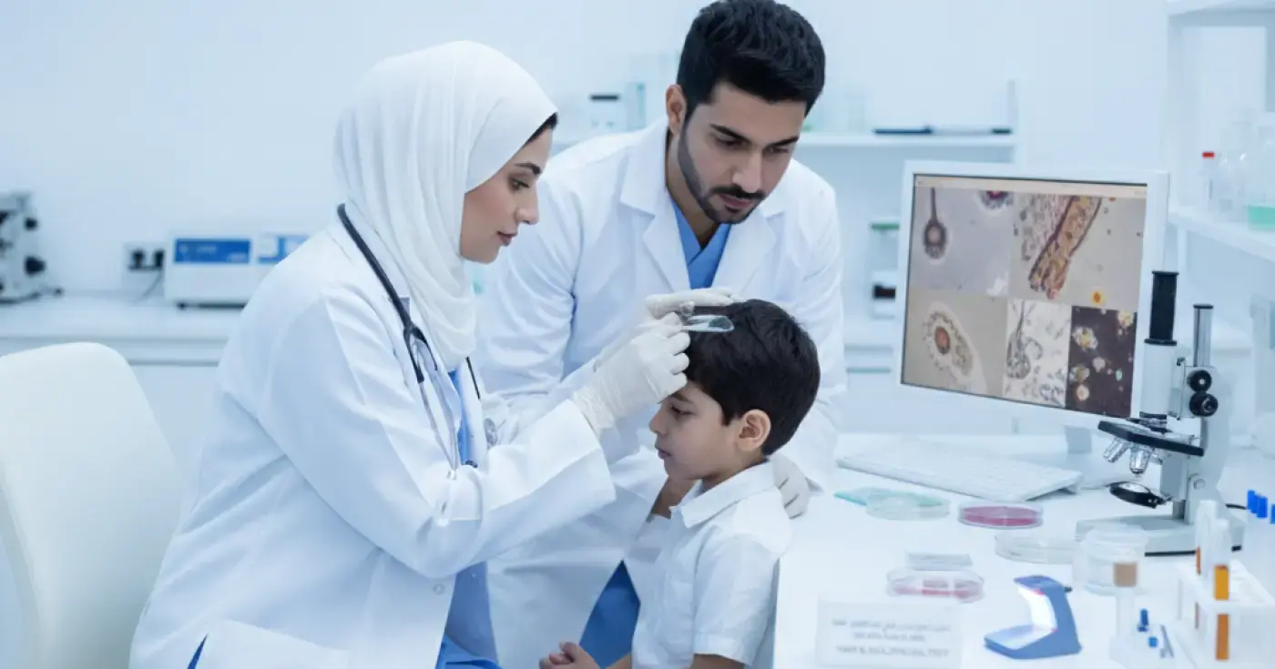
Sample collection in the laboratory is safe and minimally uncomfortable. The healthcare staff typically performs the following:
Laboratory diagnosis relies on two complementary methods:
| Test Type | Turnaround Time | Primary Objective | Procedure |
|---|---|---|---|
| Direct KOH Microscopy | Fast (hours to 24 hours) | Immediate visualization of fungal hyphae or spores | Treating sample with KOH solution and viewing under microscope |
| Fungal Culture | 2 to 4 weeks | Identifying specific fungal species and sensitivity | Incubating sample on specialized nutrient media |
A negative result indicates that fungal elements were not observed in the provided sample. However, it does not completely rule out infection if sample quantity was insufficient or if antifungal shampoos were used prior to testing. Persistent clinical signs warrant re-evaluation by the physician.
Preventing household spread involves key hygiene measures:
To protect family members, ensure the following personal items are not shared while the child is undergoing evaluation or treatment:
Parents should arrange a consultation when noticing new scaly patches or localized hair breakage on the scalp. Follow-up is also recommended if the condition shows no improvement after starting prescribed treatment.
Urgent medical evaluation is needed if severe inflammatory signs develop to prevent scarring hair loss:
Clear, direct answers
Advertisement
Continue exploring
Learn the differences between KOH direct microscopy, fungal culture, and PCR tests for onychomycosis diagnosis before starting antifungal therapy.
Read article
تعرف على الفروق بين فطريات الجلد والأكزيما والصدفية، وأهمية فحص كشط الجلد ومزرعة الفطريات للوصول للتشخيص الدقيق وتجنب العلاج العشوائي.
Read articleConnected next steps