Lahalak Medical PlatformDifferentiate Care
Learn the differences between KOH direct microscopy, fungal culture, and PCR tests for onychomycosis diagnosis before starting antifungal therapy.
Advertisement
Changes in nail color, thickness, or texture are common complaints that often lead individuals to assume an immediate diagnosis of nail fungus (Onychomycosis). However, clinical evidence shows that nearly half of all dystrophy cases stem from non-fungal etiologies, including nail psoriasis, micro-trauma, lichen planus, and circulatory disorders. Accurate diagnosis relies on combining physical examination with diagnostic lab testing to confirm the presence and species of fungi before committing to long-term treatments.
While fungi are a frequent cause of nail dystrophy, they are far from the sole cause. Changes such as yellowing, thickening, crumbling, or nail plate separation (onycholysis) occur across various dermatological condition differentials:
Empirically starting antifungal medications without diagnostic confirmation exposes patients to potential drug side effects without therapeutic benefit if the root cause is non-fungal.
Fungal nail infections are significantly more prevalent in toenails than fingernails. Clinical signs indicating the need for diagnostic sampling include:
In many patients, the infection originates from tinea pedis (athlete's foot) and gradually spreads to the nail matrix, necessitating comprehensive foot and nail evaluation.
Accurate diagnostic yield depends directly on specimen quality. Superficial clippings often lead to inconclusive findings because viable fungi reside deep within the subungual space.
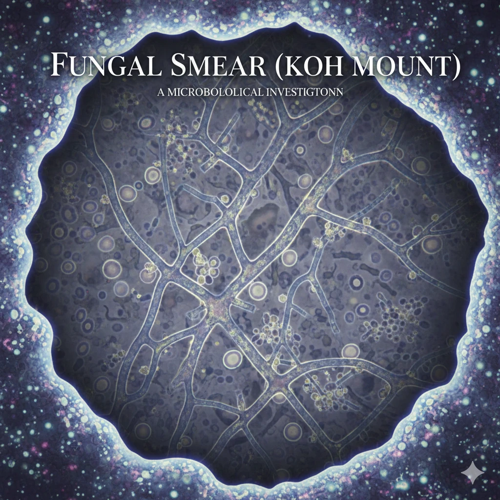
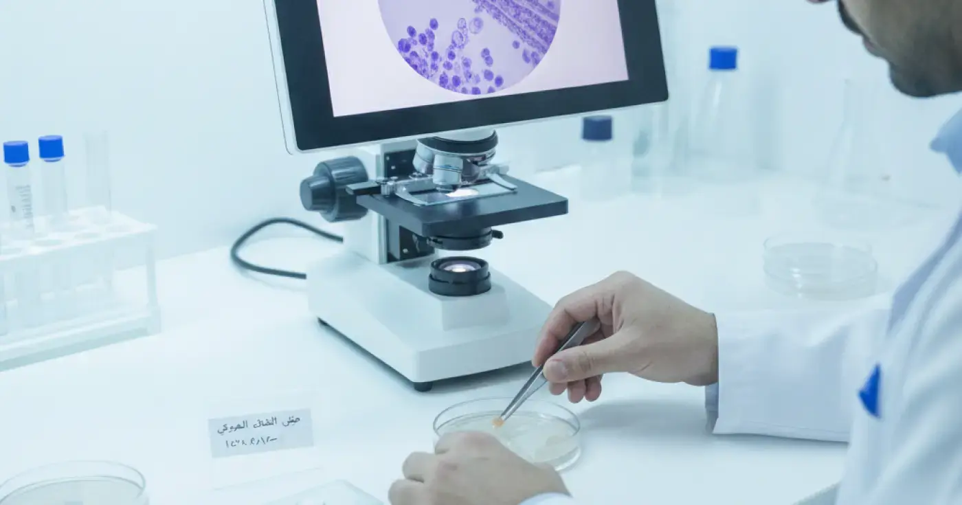
The KOH nail examination (Potassium Hydroxide wet mount) serves as the primary screening test for fungal elements. Potassium hydroxide dissolves rigid nail keratin, rendering fungal hyphae and spores visible under microscopic examination.
KOH examination provides rapid turnaround times—often within hours. While highly sensitive in detecting fungal structures, it cannot differentiate between fungal species or determine whether the organisms are viable or rendered non-viable by prior therapies.
A nail fungal culture involves inoculating nail scrapings onto specialized growth media such as Sabouraud Dextrose Agar. Cultured plates are incubated under controlled conditions and monitored weekly for colony growth.
Culture remains the standard for identifying exact fungal species, differentiating dermatophytes (e.g., Trichophyton rubrum) from non-dermatophyte molds and yeasts (e.g., Candida species). This distinction enables clinicians to select targeted antifungal regimens, particularly prior to initiating oral medications requiring liver enzyme monitoring.
The table below summarizes key differences among the three main laboratory diagnostic modalities for onychomycosis:
| Feature | Direct KOH Examination | Fungal Culture | Molecular PCR Test |
|---|---|---|---|
| Sample Type | Subungual scrapings | Sterile nail scrapings & debris | Nail clippings or scrapings |
| Primary Goal | Rapid detection of fungal hyphae | Species identification & viability | Fungal DNA sequence detection |
| Turnaround Time | Hours to 24 hours | 2 to 4 weeks | 24 to 48 hours |
| Species Identification | Low (morphology only) | High for viable cultures | Exceptionally High |
| Viability Assessment | Cannot assess viability | Detects viable organisms only | Detects intact DNA (live/dead) |
| Main Limitation | Requires a trained microscopist | Slow growth; false negatives | Higher cost & availability |
A negative KOH or fungal culture result does not completely rule out onychomycosis. Fungal culture sensitivity ranges between 60% and 70%, meaning false-negative results are relatively common due to several clinical factors:
If clinical suspicion remains high despite a negative initial result, repeat sampling, molecular PCR testing, or a nail biopsy with Periodic acid–Schiff (PAS) staining may be recommended.
Yes. Antifungal lacquers, topical creams, and oral medications suppress fungal cell growth and can inhibit culturability in the lab, leading to false-negative cultures.
Patients should discontinue topical antifungal products for at least 2 to 4 weeks before specimen collection. Oral antifungal therapy may require a longer wash-out window determined by the treating dermatologist.
Evaluation by a dermatologist ensures appropriate diagnostic selection and tailored management plans. Specialist consultation is advised when:
Fungal nail infections pose heightened health risks for individuals with diabetes, peripheral arterial disease, or compromised immune systems. Damaged nail tissue creates a portal of entry for secondary bacterial infections and cellultis.
Red flag symptoms requiring prompt medical assessment include:
To assist in choosing appropriate medical evaluation, key clinical services include:
In conclusion, thorough laboratory confirmation is the cornerstone of safe onychomycosis management, helping avoid unnecessary medications and ensuring accurate restoration of nail health.
Clear, direct answers
Advertisement