
Lahalak Medical PlatformDifferentiate Care
A clinical guide to female fertility evaluation, hormonal assays (AMH, FSH), pelvic ultrasound, and structured diagnostic protocols without over-testing.

Advertisement
Evaluating delayed conception requires a tailored clinical strategy rather than ordering exhaustive lab tests unnecessarily. Diagnostic clarity relies on combining medical history, menstrual tracking, structural imaging of the uterus and fallopian tubes, and simultaneous evaluation of the male partner.
Clinical guidelines recommend initiating a fertility workup after 12 months of regular unprotected intercourse for women under 35 years of age. For women aged 35 and older, evaluation is advised after 6 months. Patients with known risk factors, such as severe dysmenorrhea, pelvic inflammatory disease history, or irregular menstrual cycles, should seek clinical evaluation promptly.
A thorough clinical history provides essential baseline direction. Assessing cycle regularity, previous pregnancies, surgical history, and metabolic indicators helps doctors determine whether ovulatory dysfunction or structural anomalies are likely.
Ovulation can be evaluated using serum mid-luteal progesterone testing (typically measured around day 21 in a standard 28-day cycle) to confirm ovulation and corpus luteum function. Urinary luteinizing hormone (LH) detection kits can also monitor pre-ovulatory surges.
Hormonal testing evaluates ovarian reserve and baseline pituitary-ovarian signaling:
Thyroid-stimulating hormone (TSH) and prolactin screening are essential because endocrine dysregulation (such as subclinical hypothyroidism or hyperprolactinemia) can disrupt ovulation and menstrual regularity.
No. Diagnostic stewardship emphasizes selecting targeted investigations tailored to specific clinical symptoms, eliminating unnecessary costs and emotional burden.
Transvaginal ultrasound allows for pelvic examination, uterine mapping, and antral follicle count (AFC). A hysterosalpingogram (HSG) or sonohysterography assesses fallopian tube patency and uterine cavity symmetry when clinically indicated.
Male factor etiology contributes to approximately 40–50% of conception delays. A basic semen analysis evaluates sperm concentration, motility, and morphology early in the diagnostic pathway.
Maternal age influences both oocyte quantity and quality. Timely evaluation ensures appropriate counseling regarding reproductive options without unnecessary delays.
Routine unindicated screening for rare thrombophilia, advanced immunological panels, or extensive genetic screens without specific risk factors or recurrent pregnancy loss history is discouraged by international clinical guidelines.
Patients should bring past medical records, cycle tracking data, details of prior surgeries, and a list of current medications to streamline the first clinical visit.
| Clinical Question | Targeted Investigation | Routine Requirement? |
|---|---|---|
| Ovulatory status | Mid-luteal progesterone / LH kit | Yes. |
| Ovarian reserve assessment | AMH / Baseline FSH | Indicated for selected cases |
| Endocrine disruption | TSH and Prolactin | Recommended initial screen |
| Tubal factor integrity | Hysterosalpingogram (HSG) | Selected indications only |
| Male contribution | Semen analysis | Essential for all couples |
Clear, direct answers
Advertisement
Continue exploring
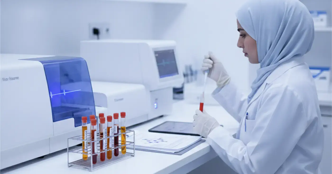
Learn about key blood tests for evaluating elevated androgen levels in women, including testosterone and DHEA-S, along with clinical evaluation steps.
Read article
A comprehensive guide on quantitative beta-hCG testing, serial measurements, ultrasound timing, and when to seek immediate medical evaluation.
Read article
Discover causes of high prolactin levels, symptoms, test preparation rules, and when doctors advise retesting for accurate diagnosis.
Read articleConnected next steps
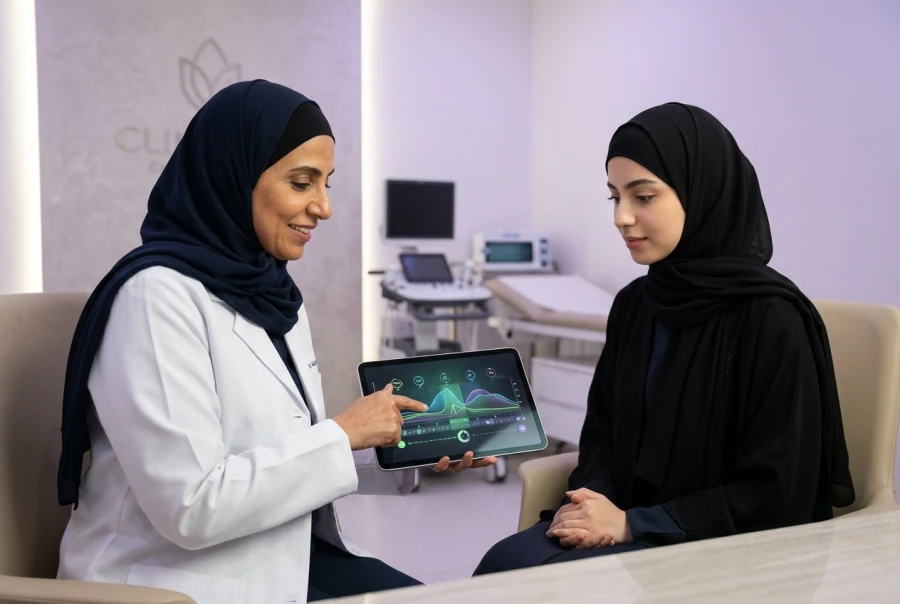 Return to Fertility Post-Contraception Education ProgramConsult our experts to understand fertility restoration after contraception. We help you plan your next pregnancy by explaining physiological changes and the expected duration for natural ovulation recovery.
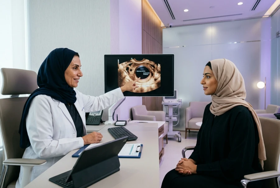 Follicular Tracking UltrasoundA precise monitoring procedure to track follicle growth and determine the optimal timing for conception, significantly enhancing pregnancy success through expert ultrasound guidance and serial development measurements.