Book OCT Corneal Epithelial Map | Lahalak Medical Platform

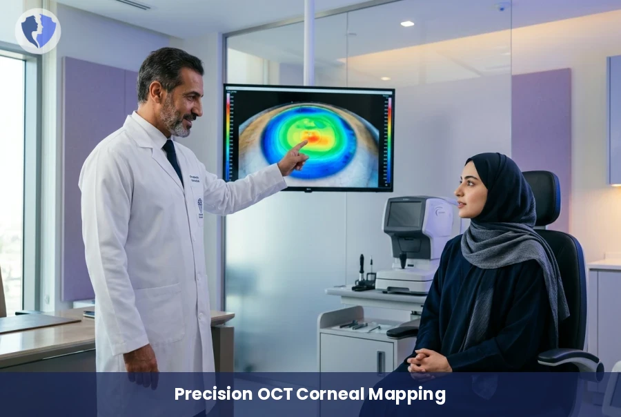
OCT Corneal Epithelial Mapping is a sophisticated diagnostic tool that measures epithelial thickness distribution, providing critical insights for managing chronic dry eye and corneal irregularities to ensure optimal visual health outcomes.

Scientific name: OCT Corneal Epithelial Mapping

A non-invasive imaging technique measuring corneal epithelial thickness distribution. It is essential for diagnosing dry eye, keratoconus, and monitoring post-refractive surgery outcomes to ensure ocular surface health and stability.

OCT Corneal Epithelial Mapping - Precision OCT Corneal Epithelial Mapping
Service type Medical Imaging
Duration 10-15 minutes
Fasting Fasting is not required

Included Services

  • OCT Corneal Scan
  • Epithelial Thickness Analysis
  • Detailed Medical Report

Medical Service Information

Why This Test is Important

  • Detects chronic dry eye conditions.
  • Early diagnosis of keratoconus.
  • Evaluates the ocular surface before and after LASIK.

Target Groups

  • Patients with chronic eye irritation.
  • Candidates for refractive surgery.
  • Keratoconus patients for regular monitoring.

Procedure Steps

  • The patient sits in front of the OCT machine.
  • Patient is asked to look at a target light.
  • Images are captured within seconds painlessly.

Conditions & Notices

  • Remove contact lenses 24 hours before the scan

Medical services you might also like