Book Cardiac MRI Iron Quantification | Lahalak Medical Platform

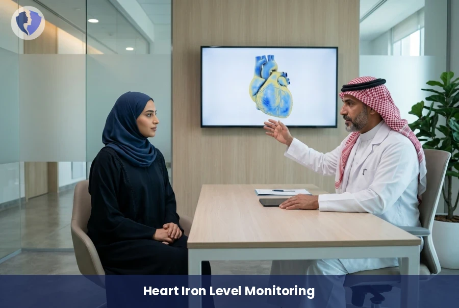
An advanced imaging technique to accurately measure iron levels in the heart muscle, helping manage chelation therapy effectively and preventing complications from iron overload for optimal cardiac health and safety.

Scientific name: MRI Cardiac Iron Quantification (T2* Mapping)

A non-invasive MRI procedure using T2* mapping to quantify iron deposition in the myocardium. Essential for thalassemia and sickle cell patients to monitor and adjust chelation therapy accurately.

MRI Cardiac Iron Quantification (T2* Mapping) - Track Your Heart Iron Levels
Service type Radiology and MRI
Duration 45-60 minutes
Fasting Fasting is not required

Included Services

  • MRI T2* Mapping Sequence
  • Detailed Medical Report
  • Iron Overload Quantification Analysis

Medical Service Information

Purpose of the Procedure

  • Early detection of iron accumulation in the heart muscle.
  • Monitoring patient response to chelation therapy.
  • Assessing the risk of heart failure due to iron overload.
Target Groups

  • Thalassemia major patients receiving frequent blood transfusions.
  • Sickle cell anemia patients.
  • Patients with Hemochromatosis conditions.

Preparation for the Test

  • No prior fasting is required for this scan.
  • All jewelry and metal objects must be removed before entering the MRI room.
  • Inform the technician if you suffer from claustrophobia.

Conditions & Notices

  • Inform the doctor of any pacemakers or metal implants
  • Bring previous medical reports and lab results

Medical services you might also like