Book 4D Pelvic Floor Ultrasound | Lahalak Medical Platform

An advanced imaging technique providing comprehensive visualization of pelvic floor muscles and tissues, essential for diagnosing prolapse and functional disorders with extreme precision to ensure the best treatment plan.

Scientific name: Advanced 3D/4D Pelvic Floor Ultrasound

A non-invasive procedure using 4D ultrasound to evaluate pelvic floor muscles, detect muscle defects, and verify the positioning of mid-urethral slings with high clinical accuracy.

Advanced 3D/4D Pelvic Floor Ultrasound - High-Precision Pelvic Floor Imaging
Service type Ultrasound Imaging
Duration 30-45 minutes
Fasting Fasting is not required

Included Services

  • 3D Pelvic Imaging
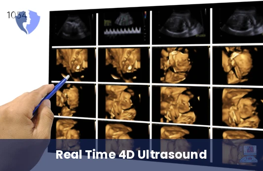
  • 4D Pelvic Imaging
  • Detailed medical report

Medical Service Information

Purpose of the Procedure

  • Evaluating the integrity of pelvic floor muscles and tissues.
  • Accurately diagnosing pelvic organ prolapse.
  • Ensuring correct positioning of mid-urethral slings.
  • Identifying muscle defects following difficult deliveries.

Target Groups

  • Women suffering from stress urinary incontinence.
  • Cases of uterine or bladder prolapse.
  • Patients who have undergone pelvic sling surgery.
  • Women experiencing chronic pelvic pain.

Advantages of 4D Imaging

  • Provides real-time visualization of muscle movement during contraction.
  • Allows for precise measurement of pelvic hiatus dimensions.
  • Assists in accurate surgical planning for corrective procedures.
  • Completely safe procedure with no radiation exposure.

Conditions & Notices

  • It is preferred to empty the bladder immediately before the scan unless otherwise directed

Medical services you might also like