Book 3D Pelvic Floor Ultrasound | Lahalak Medical Platform

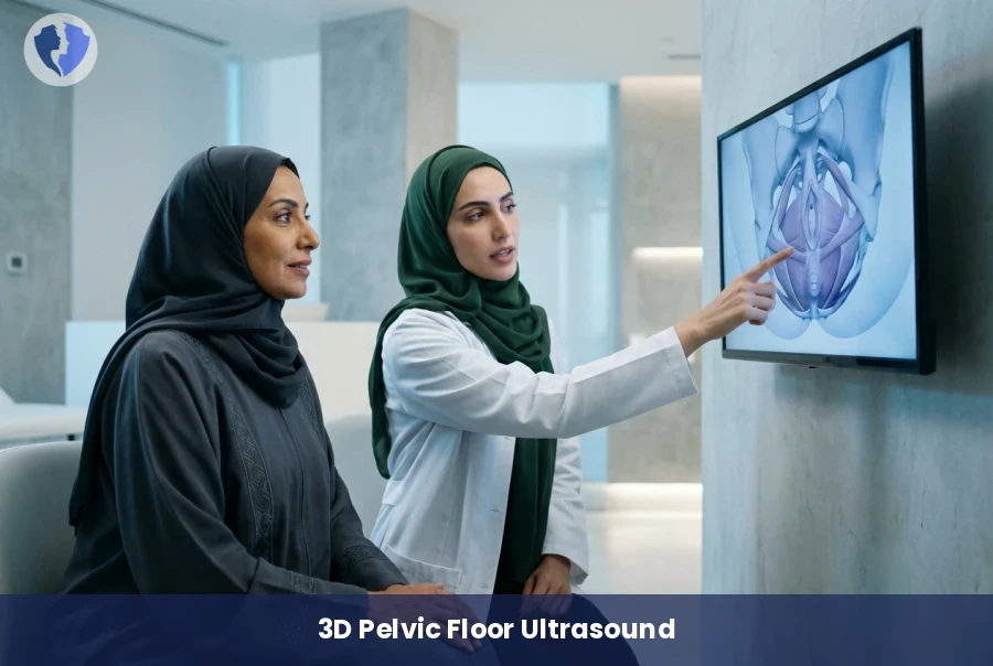
A cutting-edge 3D imaging technology designed to evaluate the integrity of pelvic support muscles, detecting levator ani avulsions and structural defects to ensure effective treatment for various pelvic floor disorders.

Scientific name: 3D Pelvic Floor Ultrasound (Levator Ani Assessment)

A non-invasive diagnostic procedure using advanced 3D ultrasound to assess the levator ani muscle. It identifies muscle avulsions or structural defects in the pelvic floor caused by childbirth or aging.

3D Pelvic Floor Ultrasound (Levator Ani Assessment) - Advanced Pelvic Floor Assessment
Service type Radiology (Ultrasound)
Duration 20-30 minutes
Fasting Fasting is not required

Included Services

  • 3D Pelvic floor imaging
  • Detailed medical report on muscle status
  • Medical consultation to discuss results

Medical Service Information

Purpose of the Procedure

  • Assess the integrity of the levator ani muscle.
  • Detect muscle avulsions resulting from vaginal delivery.
  • Diagnose causes of pelvic organ prolapse and urinary incontinence.
Target Groups

  • Women experiencing symptoms of vaginal or uterine prolapse.
  • Postpartum women after difficult vaginal births to assess muscle damage.
  • Menopausal women with bladder control issues.

Technique Advantages

  • High precision in soft tissue imaging compared to traditional X-rays.
  • Completely safe procedure with no harmful radiation.
  • Provides a comprehensive view of pelvic angles difficult to reach with standard ultrasound.

Conditions & Notices

  • It is preferred to empty the bladder immediately before the scan

Medical services you might also like