
Lahalak Medical PlatformDifferentiate Care
Learn how dermatologists evaluate moles using dermoscopy, visual exams, and skin biopsies to ensure early detection of atypical lesions.
Advertisement
Moles (nevi) are common skin growths that most people develop during their lifetime. While the vast majority of moles are benign and harmless, some can undergo atypical changes that require professional medical evaluation to rule out skin cancers, such as melanoma. Regular clinical mole evaluation plays a vital role in early detection, significantly improving treatment outcomes and patient safety.
Dermatologists recommend performing a monthly skin self-exam at home. You should schedule a clinical evaluation immediately if you notice any changes following the ABCDE rule:
During your visit to a dermatology clinic, the specialist follows a structured, scientific approach to assess your moles. This process typically includes:
The dermatologist begins by discussing your personal and family medical history, focusing on any history of skin cancer, sun exposure habits, history of sunburns, and any specific moles that have recently changed or caused concern.
The doctor performs a full-body skin exam from head to toe, including easily overlooked areas such as the scalp, behind the ears, between the toes, and under the nails. This helps identify any "ugly duckling" lesions—moles that look significantly different from the patient's other moles.
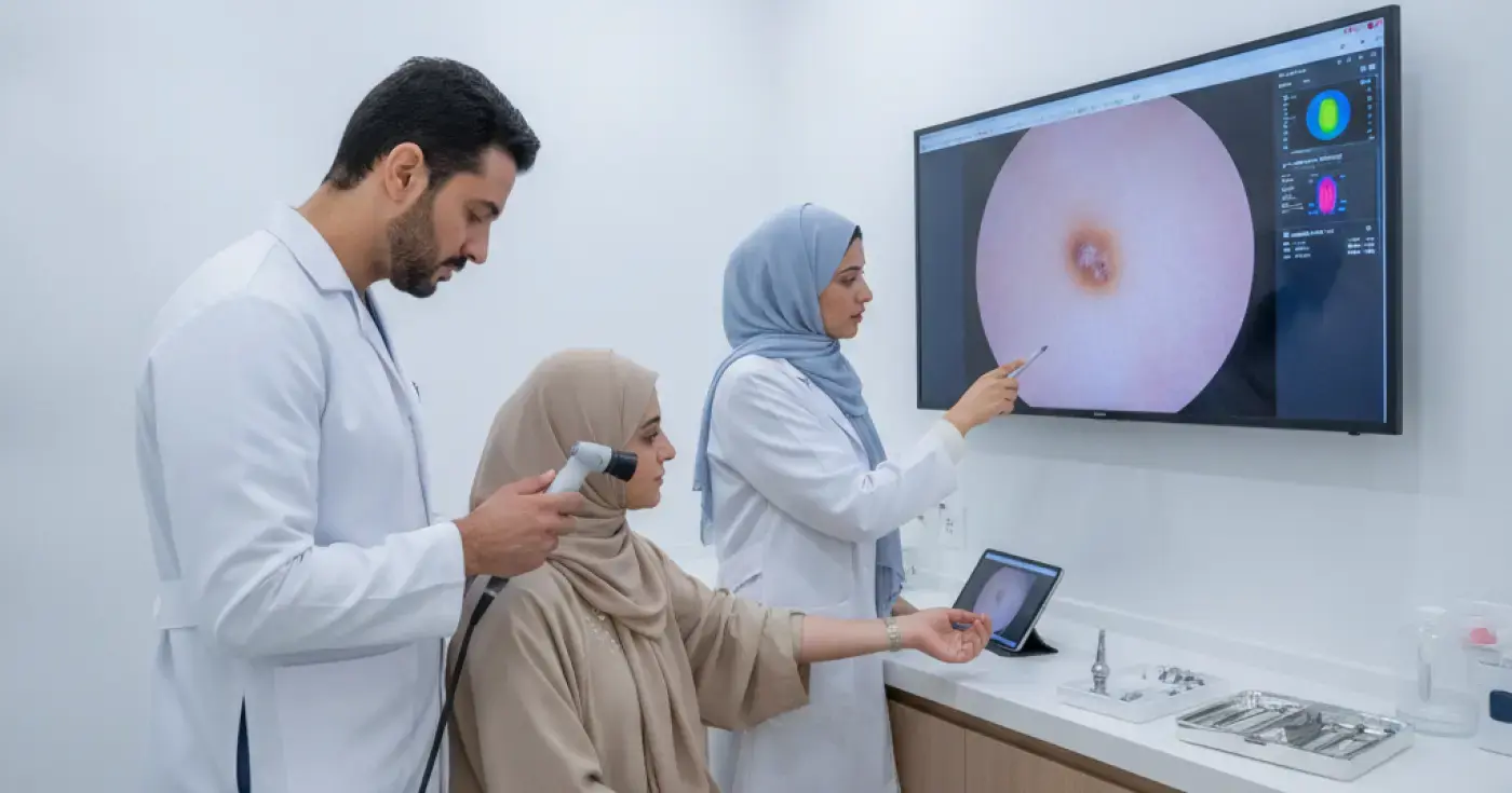
The dermatologist uses a specialized handheld tool called a dermatoscope. This device provides high magnification and cross-polarized lighting, allowing the physician to visualize pigment patterns and deep microstructures within the skin layers that are invisible to the naked eye. This non-invasive step highly increases diagnostic accuracy.
If a mole displays suspicious features during the visual or dermoscopic exam, advanced diagnostic steps may be recommended:
For patients with a high number of atypical moles (often more than 50), dermatologists may use total body photography. High-resolution baseline photos are taken and stored to track and compare any subtle changes or new lesions during follow-up visits.
If a mole is highly suspicious, the dermatologist will perform a skin biopsy. Under local anesthesia, the doctor removes the entire mole or a sample of it. The tissue is sent to a pathology lab where a dermatopathologist examines the cells under a microscope to provide a definitive diagnosis.
The following table highlights the key clinical differences evaluated by dermatologists:
| Clinical Feature | Benign Mole (Common Nevus) | Atypical Mole (Dysplastic Nevus) |
|---|---|---|
| Shape | Symmetrical, round, or oval | Asymmetrical (halves do not match) |
| Borders | Sharp, smooth, and well-defined | Irregular, notched, or blurred |
| Color | Uniform single shade of brown or tan | Varied shades of brown, black, red, or pink |
| Size | Usually less than 6 mm | Often larger than 6 mm and may grow |
| Symptoms | Asymptomatic (no symptoms) | May itch, hurt, crust, or bleed |
Based on the clinical findings, the dermatologist will recommend the next steps:
To protect your skin and maintain long-term health, follow these essential guidelines:
Clear, direct answers
Advertisement
Continue exploring

Learn the importance of regular mole screening, the ABCDE rule for self-exams, and warning signs that require an immediate visit to a dermatologist.
Read article
Discover how many sessions are required for a thread lift, how long the results last, and the key factors influencing your treatment plan.
Read article
Learn about normal thread lift recovery, potential side effects, and warning signs that require immediate medical consultation.
Read article