Leishmania Detection Smear | Lahalak Medical Platform
Direct and accurate diagnosis of Leishmania via microscopic examination of tissue samples. This ensures immediate start of correct treatment to prevent skin or visceral infection spread.
Scientific name: Leishmania Detection Smear
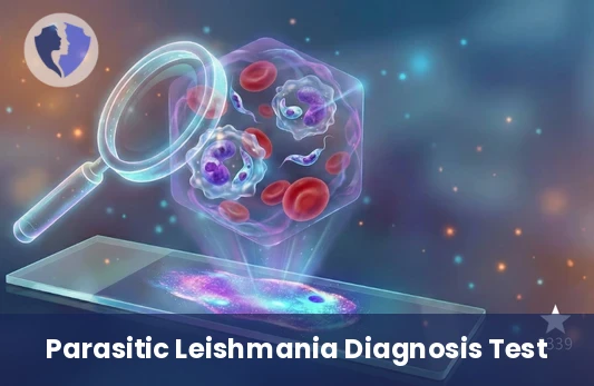
Direct microscopic examination of a sample (from skin lesion, bone marrow, lymph node, or spleen) after staining with Giemsa stain to search for intracellular parasite form (amastigote) specific to Leishmania. Considered one of the primary direct diagnostic methods.
Service type
Microscopic examination (Tissue smear)
Duration
15-20 minutes
Fasting
Fasting is not required
Included Services
- Direct microscopic examination to detect the Leishmania parasite in samples.
- Consult an infectious disease or dermatology specialist to interpret the results.
- Personal support services from Halak.
Medical Service Information
Conditions & Notices
- Fasting: Not required.
- Sample collection: The sample should be taken from the active edge of the ulcer, not from the dead or suppurating center. It should be taken under sterile conditions.
- Sample type: According to disease type:
- Dermatological: A swab or biopsy from the edge of the ulcer.
- Visceral: A sample of bone marrow (often sternum) or spleen (more accurate but dangerous).
- Transportation: The sample must be sent to the laboratory quickly. It can be placed in a special transport medium if the analysis is delayed.
Medical services you might also like