Service Details
Renal Light Microscopy Biopsy - Renal Biopsy: Light Microscopy with Special Stains
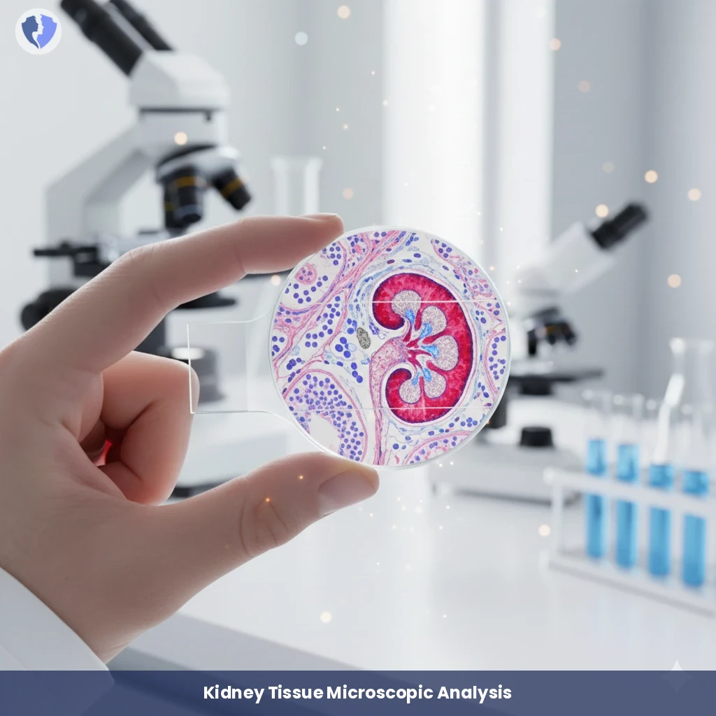
Histopathological examination of a kidney biopsy specimen using a light microscope with the application of a range of specialized histological stains to paraffin-encapsulated tissue slides. Common stains include: H&E (hematoxylin and eosin) for general evaluation, PAS (periodic acid Schiff) for staining the glomerular basement membrane and matrix, Trichrome (Mason) for differentiating collagen (fibrosis/scarring) from other tissues, and Jones silver stain for detailing the glomerular basement membrane. The examination aims to diagnose glomerulonephritis, sclerosis, and tubulointerstitial changes.